
Page 1829 - Hall et al (2015) Principles of Critical Care-McGraw-Hill
P. 1829
1298 PART 11: Special Problems in Critical Care
of cleavage. Pathologic findings are outlined in Table 129-11. Biopsies
at the border of a blister and normal skin should also be sent for routine
processing. Cultures from blisters are negative because of the toxin-
mediated nature of the disease.
Treatment of SSSS requires appropriate antibiotics and careful moni-
toring of fluids and electrolytes. An antibiotic with activity against
β-lactamase–producing S aureus is recommended. Topical antibiotics
are not necessary. Adjunctive measures include the application of bland
lubricants for the patient’s comfort. Healing occurs in 7 to 10 days.
■ PSEUDOMONAS BACTEREMIA
Several skin manifestations of Pseudomonas bacteremia have been
described, and fall into four general categories. The first is ecthyma
108
gangrenosum (Fig. 129-28), which is characterized by a localized, ery-
thematous, tender plaque or bulla that subsequently develops central
necrosis, leaving a gangrenous eschar with an erythematous annular
border. The lesions can occur anywhere but are usually found in the
anogenital region, buttocks, or axilla. Ecthyma gangrenosum occurs
109
in approximately 5% of patients with Pseudomonas bacteremia and has
been described in association with localized Pseudomonas infection
without bacteremia. The second category includes vesicles or bullae that
can occur anywhere and may occur singly or in clusters. These lesions
frequently become hemorrhagic and take on the appearance of ecthyma
gangrenosum lesions when ruptured. The third category is cellulitis with
a sharply demarcated border, unlike cellulitis caused by staphylococ-
cal or streptococcal infection, which tends to have ill-defined borders.
The fourth category of lesions comprises small pink, round plaques or
subcutaneous nodules that are concentrated on the trunk and proximal
extremities. The nodules are considered a form of nodular cellulitis and,
when incised and drained, grow Pseudomonas aeruginosa in culture.
Pseudomonas aeruginosa bacteremia carries a high mortality rate and
appropriate antimicrobial treatment should be initiated early.
■ MENINGOCOCCEMIA
Acute infection with the gram-negative diplococcus Neisseria meningiti-
dis is associated with characteristic cutaneous findings, which may aid
in the early diagnosis of this rapidly fatal disease. Cutaneous findings
are present in more than 70% of meningococcemia cases. Characteristic
findings include petechiae, ecchymoses, and palpable purpura. Petechiae
may have a smudged appearance and tend to be concentrated on the
trunk, proximal extremities, and mucosal surfaces (Fig. 129-29). The
number of petechiae correlates with the degree of thrombocytopenia
and is indistinguishable from other causes of petechiae, such as diffuse
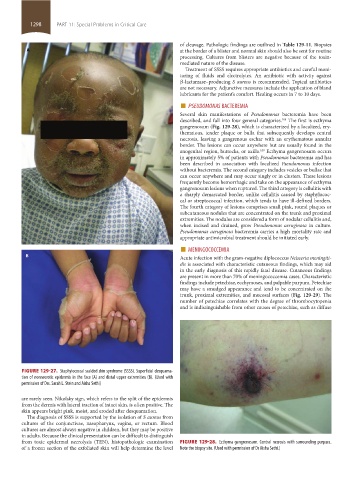
FIGURE 129-27. Staphylococcal scalded skin syndrome (SSSS). Superficial desquama-
tion of nonnecrotic epidermis in the face (A) and distal upper extremities (B). (Used with
permission of Drs. Sarah L. Stein and Aisha Sethi)
are rarely seen. Nikolsky sign, which refers to the split of the epidermis
from the dermis with lateral traction of intact skin, is often positive. The
skin appears bright pink, moist, and eroded after desquamation.
The diagnosis of SSSS is supported by the isolation of S aureus from
cultures of the conjunctivae, nasopharynx, vagina, or rectum. Blood
cultures are almost always negative in children, but they may be positive
in adults. Because the clinical presentation can be difficult to distinguish
from toxic epidermal necrolysis (TEN), histopathologic examination FIGURE 129-28. Ecthyma gangrenosum. Central necrosis with surrounding purpura.
of a frozen section of the exfoliated skin will help determine the level Note the biopsy site. (Used with permission of Dr Aisha Sethi.)
section11.indd 1298 1/19/2015 10:55:27 AM