
Page 1826 - Hall et al (2015) Principles of Critical Care-McGraw-Hill
P. 1826
CHAPTER 129: Dermatologic Conditions 1295
Palpable purpura
No systemic involvement With systemic involvement
(primary cutaneous vasculitis) (systemic vasculitis)
I. Leukocytoclastic vasculitis *GI involvement Fever
1. Idiopathic (bloody stools)
2. Drug-induced *Renal involvement
(NSAIDs, penicillin, quinolones, (hematuria) * Rule out underlying
minocycline, hydralazine, anti- *Arthritis infection vs malignancy
TNF- agents) * +/– asthma
-> Bacterial/viral/fungal
cultures
Cryo + -> Hep B/C, CMV, HIV,
ANCA – parvovirus B19
-> Echo if heart murmur
Cryo –
ANCA +
ANCA –
I. Wegener granulomatosis I. Cryoglobulinemic I. Rheumatoid vasculitis
(c-ANCA) vasculitis II. Sjögren disease
II. Churg-Strauss (p-ANCA) -> Rule out underlying III. Systemic lupus
III. Microscopic polyangiitis multiple myeloma, other IV. Urticarial vasculitis
lymphoproliferative V. Henoch-Schönlein purpura
disorders
-> HCV, HBV, HIV
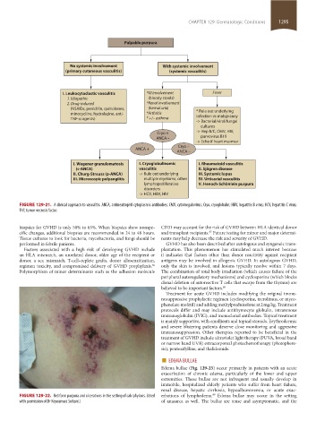
FIGURE 129-21. A clinical approach to vasculitis. ANCA, antineutrophil cytoplasmic antibodies; CMV, cytomegalovirus; Cryo, cryoglobulin; HBV, hepatitis B virus; HCV, hepatitis C virus;
TNF, tumor necrosis factor.
biopsies for GVHD is only 50% to 65%. When biopsies show nonspe- CD31 may account for the risk of GVHD between HLA identical donor
cific changes, additional biopsies are recommended in 24 to 48 hours. and transplant recipients. Future testing for minor and major determi-
87
Tissue cultures to look for bacteria, mycobacteria, and fungi should be nants may help decrease the risk and severity of GVHD.
performed in febrile patients. GVHD has also been described after autologous and syngeneic trans-
Factors associated with a high risk of developing GVHD include plantation. This phenomenon has stimulated much interest because
an HLA mismatch, an unrelated donor, older age of the recipient or it indicates that factors other than donor reactivity against recipient
donor, a sex mismatch, T-cell–replete grafts, donor allosensitization, antigens may be involved in allogenic GVHD. In autologous GVHD,
regimen toxicity, and compromised delivery of GVHD prophylaxis. only the skin is involved, and lesions typically resolve within 7 days.
86
Polymorphism of minor determinants such as the adhesion molecule The combination of total body irradiation (which causes failure of the
peripheral autoregulatory mechanisms) and cyclosporine (which blocks
clonal deletion of autoreactive T cells that escape from the thymus) are
believed to be important factors. 88
Treatment for acute GVHD includes modifying the original immu-
nosuppressive prophylactic regimen (cyclosporine, tacrolimus, or myco-
phenolate mofetil) and adding methylprednisolone at 2 mg/kg. Treatment
protocols differ and may include antithymocyte globulin, intravenous
immunoglobulin (IVIG), and monoclonal antibodies. Topical treatment
is mainly supportive, with emollients and topical steroids. Erythrodermic
and severe blistering patients deserve close monitoring and aggressive
immunosuppression. Other therapies reported to be beneficial in the
treatment of GVHD include ultraviolet light therapy (PUVA, broad band
or narrow band UVB) extracorporeal photochemotherapy (photophere-
sis), pentoxifylline, and thalidomide.
■ EDEMA BULLAE
Edema bullae (Fig. 129-25) occur primarily in patients with an acute
exacerbation of chronic edema, particularly of the lower and upper
extremities. These bullae are not infrequent and usually develop in
immobile, hospitalized elderly patients who suffer from heart failure,
renal disease, hepatic cirrhosis, hypoalbuminemia, or acute exac-
89
FIGURE 129-22. Retiform purpura and ulcerations in the setting of calciphylaxis. (Used erbations of lymphedema. Edema bullae may occur in the setting
with permission of Dr Keyoumars Soltani.) of anasarca as well. The bullae are tense and asymptomatic, and the
section11.indd 1295 1/19/2015 10:54:23 AM