
Page 1820 - Hall et al (2015) Principles of Critical Care-McGraw-Hill
P. 1820
CHAPTER 129: Dermatologic Conditions 1289
acetate, and ophthalmic sodium sulfacetamide) because of potential
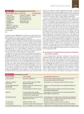
TABLE 129-10 Clinical and Histopathological Features of SJS/TEN
significant absorption and the possibility of inducing SJS. A variety of
54
Laboratory Findings (% patients) Histopathology Immunofluorescence other dressings may be used to decrease fluid loss and pain and promote
55
• Anemia (100%) • Subepidermal vesiculation Negative wound healing. These include porcine xenografts and synthetic dress-
• Lymphopenia (90%) • Necrotic keratinocytes at all ings such as Omniderm, OpSite, Vigilon, Mepitel, and Biobrane.
• Neutropenia (30%) levels of the epidermis in SJS The use of corticosteroids and immunosuppressive agents in the
a
• Thrombocytopenia b • Full-thickness necrosis in TEN treatment of TEN is controversial. The rationale for such an approach is
• Eosinophilia • Minimal inflammatory based on the assumption that TEN is an immune-mediated event. There
• Hypoalbuminemia infiltrate in the dermis are no case-controlled studies supporting the use of corticosteroids;
• Hypocalcemia however, several case reports have advocated their use. 56,57 In most cases
• Proteinuria, <1 g/day (50%) in which steroids were found effective, they were started very early in
• Elevated transaminases (30%) the course of the disease. More recent literature has indicated a higher
• Elevated amylase, lipase mortality rate, increased time to recovery, and increased length of hos-
pital stay in patients treated with systemic corticosteroids. 58,59 Patients
a Carries poor prognosis.
undergoing long-term glucocorticoid therapy who develop TEN may
b Rare. have a delay in onset, but the severity of disease is unaffected. Based
60
on this information, corticosteroids should not be used routinely in
pemphigus vulgaris (Table 129-11). In all these cases, full-thickness epi- TEN and should only be considered in patients who present early in the
dermal necrosis is rare (except in GVHD, in which epidermal necrosis is course of disease. Intravenous immunoglobulin therapy (IVIG) in TEN
accompanied by an abundant lymphocytic infiltrate). aims to decrease Fas-mediated keratinocyte apoptosis by sequestering
Erythema multiforme (Fig. 129-13) (previously referred to as erythema Fas available for binding to CD95. Several nonrandomized, uncontrolled
multiforme minor) is an acute, self-limited reaction characterized by studies have shown that large-dose IVIG decreases mortality rate and
asymptomatic, annular erythematous, or urticarial plaques with central progression of TEN, 61-64 whereas at least one prospective study showed
areas of blistering and necrosis, resulting in the characteristic target no benefit. Currently, there is no consensus about the use of IVIG due
40
lesions. Lesions usually develop on the extensor surfaces of the extremi- to lack of controlled, randomized trials. In addition, the use of plasma-
ties and mucosae, and cover less than 10% of the body surface area. pheresis and immunosuppressive drugs remains controversial due to the
Outbreaks last for 1 to 4 weeks. Relapses are frequent. It is associated lack of strong clinical data.
with recurrent herpes simplex virus infections in the great majority of
Treatment for SJS and TEN includes pain management and prompt ■
cases and with M pneumoniae infection in a smaller subset. HYPERSENSITIVITY SYNDROME/DRUG REACTION WITH EOSINOPHILIA
withdrawal of any agents that are not essential for the maintenance of AND SYSTEMIC SYMPTOMS
life (Table 129-12). The latter has been shown to decrease mortality rate A severe hypersensitivity syndrome consisting of fever, skin rash,
in TEN by about 30% per day, despite some progression of the muco- lymphadenopathy, and variable organ (usually liver) involvement may
cutaneous involvement. Fluid resuscitation, treatment of infections, appear 1 to 8 weeks after administration of the inciting drug for the first
42
and meticulous skin and eye care are most appropriately managed in an time. When associated with eosinophilia and systemic symptoms, the
isolation room in a burn unit, where the staff is trained in topical wound term drug reaction with eosinophilia and systemic symptoms (DRESS)
and skin care. Topical therapy includes gentle debridement of necrotic is used. Table 129-13 includes the most recently proposed diagnostic
skin followed by the application of nonadherent dressings to the areas criteria for DRESS. Typical precipitating drugs are aromatic anticon-
65
of skin erosion, a wrapping of a thin silver-impregnated dressing, and vulsants (eg, phenytoin, phenobarbital, and carbamazepine) and sulfon-
an outer linen covering to hold all in place. Antibiotic ointments are amides. DRESS syndrome is estimated to occur in between 1 in 1000
widely used, although their value is unproven. It is important to avoid and 1 in 10,000 exposures with these drugs. There is a 75% incidence
the sulfonamide derivatives (silver sulfadiazine cream, topical mafenide of cross-reactivity between the different anticonvulsants; therefore,
TABLE 129-11 Differential Diagnosis of SJS/TEN
Differential Diagnosis Cutaneous Findings Histopathology/Immunofluorescence
Erythema multiforme Asymptomatic, targetoid lesions Interface dermatitis with individual apoptotic keratinocytes and a perivascular
lymphocytic infiltrate/negative immunofluorescence
Staphylococcal scalded skin Large areas of tender erythema, flaccid bullae Subcorneal blisters with no inflammatory cells/immunofluorescence negative
syndrome (SSSS) followed by desquamation
Acute generalized exanthematous Nonfollicular, sterile pustules within large areas of Subcorneal or superficial epidermal blisters with neutrophils/immunofluorescence
pustulosis (AGEP) edematous erythema negative
Graft-versus-host disease Generalized erythematous morbilliform eruption Vacuolar interface dermatitis with satellite necrotic keratinocytes and epithelial
atypia/negative immunofluorescence
Scarlet fever Erythematous papules on the trunk with a Engorged capillaries and dilated lymphatic vessels, most prominent around hair
“ sandpaper-like” texture followed by desquamation follicles/negative immunofluorescence
of the hands and feet, strawberry tongue
Paraneoplastic pemphigus Mucocutaneous involvement with tense and flaccid Variable intraepidermal acantholysis, interface reaction with necrotic keratinocytes
bullae intermixed with erosions. and vacuolar change, +/− subepidermal clefting/ IgG and C3 deposition between
keratinocytes and at the dermoepidermal junction
Pemphigus vulgaris Superficial flaccid bullae, erosions Intraepidermal split/IgG and C3 deposition between keratinocytes
Bullous pemphigoid Large tense bullae, urticarial wheals, serpiginous plaques Subepidermal blister/linear deposition of IgG and C3 along basement membrane
Drug-induced linear IgA bullous dermatosis Tense vesicles and bullae with annular configuration Subepidermal blister with neutrophils/linear deposition of IgA along the dermoepidermal junction
section11.indd 1289 1/19/2015 10:53:22 AM