
Page 617 - Clinical Hematology_ Theory _ Procedures ( PDFDrive )
P. 617
CHAPTER 29 ■ Body Fluid Analysis 601
o demonstrating pericardial e usion and are less subject to ( microhematocrit), however, can be valuable in distin-
the limitations o echocardiography in localized e usions. guishing a hemorrhagic e usion rom aspirated blood in a
Pericardial disease causes e usions that are le sided or specimen. T e quantity o erythrocytes is usually lower in a
bilateral; they are rarely exclusively right sided. Patients with hemorrhagic e usion than in a simultaneously assayed cir-
congestive heart ailure typically mani est right-sided or culating blood specimen. In contrast, aspirated blood, i su -
bilateral e usions. cient in quantity, will exhibit an erythrocyte volume that is
comparable to that in the circulating blood.
Causes of Pericardial Effusion Pericardial f uid is relatively acellular. An increase (more
A wide variety o diseases and disorders can produce peri- than 1 × 10 /L) is suggestive o microbial in ection or
9
cardial e usion ( able 29.11). Neoplastic disease pro- malignancy.
duces a signi cant volume o f uid in the pericardium and
is one o the most common causes o pericardial e usion. Evaluation of Sm ears
Primary tumors o the pericardium (mesothelioma) are Sediment should be prepared or microscopic examination
rare. However, metastatic tumors o the pericardium and as previously described in the section “Pleural Fluid.” T e
heart are common in patients with advanced malignant dis- sediment should be stained and examined or leukocytic
ease rom primary sites (such as the lung and breast) and in cells and malignant mesothelial cells.
patients with leukemia or lymphoma. T ese types o metas-
tases are the most common causes o malignant e usions. Leukocyte Dif erential
T ere ore, one o the most important parts o the labora- Te value o a di erential leukocyte count in establishing a
tory examination o pericardial f uid is cytological studies di erential diagnosis is debatable. However, an elevated total
or malignant cells. leukocyte count in conjunction with mostly PMNs can be
observed in bacterial pericarditis. In contrast, pericardial f uid
Laboratory Analysis may demonstrate increased lymphocytes in viral pericarditis.
Gross Exam ination Mononuclear phagocytes (monocytes, histiocytes, and
Normal f uid is transparent and pale yellow. Hemorrhagic macrophages) can be seen in variable numbers in pericar-
(bloody) e usions may result rom a variety o abnormal dial e usions. In addition, in vivo LE cell ormation has been
conditions or rom aspiration o intracardiac blood into the observed in pericardial f uids.
specimen. On visible examination, a hemorrhagic e usion
should not orm clots in a plain (nonanticoagulant) tube, but Cytological Examination
aspirated blood usually exhibits clotting. A milky-appearing Smears should be closely examined or the presence o malig-
e usion may be a true or pseudochylous f uid (see “Pleural nant mesothelial cells. T e appearance o these cells was pre-
Fluid” or a discussion o milky e usions). viously described in “Pleural Fluids.”
T e value o the measurement o pH is not well estab-
lished. However, specimens with a pH less than 7.0 may be NOTE: This is a good time to complete Review Questions
associated with in ectious or rheumatoid disease. In addi- related to the preceding content.
tion, hemorrhagic specimens typically demonstrate a pH
that is lower than the pH in circulating blood.
Seminal Fluid
Cell Counts T e main unction o seminal f uid (semen) is to transport
Erythrocyte and leukocyte cell counts are o limited value sperm to emale cervical mucus. A er deposition in the
in the di erential diagnosis o a pericardial e usion. emale reproductive tract, sperm remain in the seminal
Erythrocyte counts or a determination o packed cell volume plasma or a short time while attempting to enter the mucus.
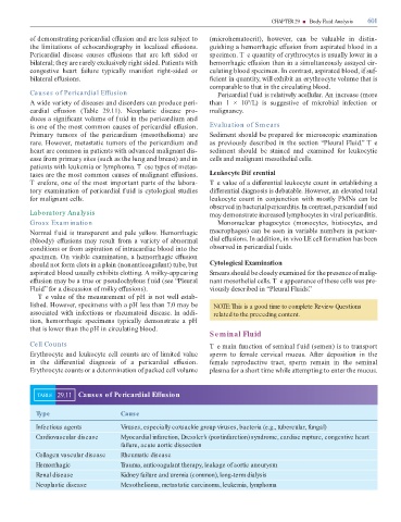
TABLE 29.11 Causes of Pericardial Effusion
Type Cause
Infectious agents Viruses, especially coxsackie group viruses, bacteria (e.g., tubercular, fungal)
Cardiovascular disease Myocardial infarction, Dressler’s (postinfarction) syndrome, cardiac rupture, congestive heart
failure, acute aortic dissection
Collagen vascular disease Rheumatic disease
Hemorrhagic Trauma, anticoagulant therapy, leakage of aortic aneurysm
Renal disease Kidney failure and uremia (common), long-term dialysis
Neoplastic disease Mesothelioma, metastatic carcinoma, leukemia, lymphoma