
Page 654 - Clinical Hematology_ Theory _ Procedures ( PDFDrive )
P. 654
638 PART 8 ■ Fundamentals of Hematological Analysis
More recently, monoclonal antibodies directed against f uorescence measurements. T e cell type o interest can be
CD20, CD25, CD33, CD45, and CD52 have been developed. separated rom a complex mixture o cell types even though
Be ore treatment, f ow-cell analysis is critical or con rming it may be an extremely rare or minor subpopulation.
that the antigen is expressed by the o ending cells. During
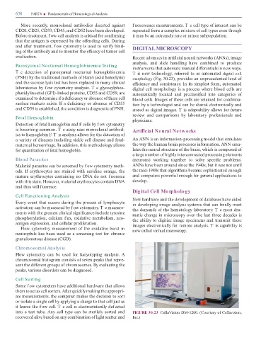
and a er treatment, f ow cytometry is used to veri y bind- DIGITAL MICROSCOPY
ing o the antibody and to monitor the e cacy o tumor cell
eradication. Recent advances in arti cial neural networks (ANNs), image
analysis, and slide handling have combined to produce
Paroxysm al Nocturnal Hem oglobinem ia Testing
instruments that automate manual di erentials in new ways.
T e detection o paroxysmal nocturnal hemoglobinemia T is new technology, re erred to as automated digital cell
(PNH) by the traditional methods o Ham’s (acid hemolysis) morphology (Fig. 30.23), provides an unprecedented level o
and the sucrose lysis test has been replaced in many clinical e ciency and consistency. In its simplest orm, automated
laboratories by f ow cytometry analysis. T e glycosylphos- digital cell morphology is a process where blood cells are
phatidylinositol (GPI)-linked proteins, CD55 and CD59, are automatically located and preclassi ed into categories o
examined to determine i a de ciency or absence o these cell blood cells. Images o these cells are retained or con rma-
sur ace markers exists. I a de ciency or absence o CD55 tion by a technologist and can be shared electronically and
and CD59 is established, the condition is diagnostic o PNH. stored as digital images. T is adaptability allows or uture
review and comparisons by laboratory pro essionals and
Fetal Hem oglobin
physicians.
Detection o etal hemoglobin and F cells by f ow cytometry
is becoming common. T e assay uses monoclonal antibod- Arti cial Neural Networks
ies to hemoglobin F. T is analysis allows or the detection o
a variety o diseases including sickle cell disease and etal- An ANN is an in ormation-processing model that simulates
maternal hemorrhage. In addition, this methodology allows the way the human brain processes in ormation. ANN emu-
or quantitation o etal hemoglobin. lates the neural structure o the brain, which is composed o
a large number o highly interconnected processing elements
Blood Parasites (neurons) working together to solve speci c problems.
Malarial parasites can be screened by f ow cytometry meth- ANNs have been around since the 1940s, but it was not until
ods. I erythrocytes are stained with acridine orange, the the mid-1980s that algorithms became sophisticated enough
mature erythrocytes containing no DNA do not f uoresce and computers power ul enough or general applications to
with this stain. However, malarial erythrocytes contain DNA develop.
and thus will f uoresce.
Digital Cell Morphology
Cell Functioning Analysis
New hardware and the development o databases have aided
Every event that occurs during the process o lymphocyte in developing image analysis systems that can nally meet
activation can be measured by f ow cytometry. T e measure- the demands o the hematology laboratory. T e most dra-
ments with the greatest clinical signi cance include tyrosine matic change in microscopy over the last three decades is
phosphorylation, calcium f ux, oxidative metabolism, neo- the ability to digitize image specimens and transmit these
antigen expression, and cellular proli eration. images electronically or remote analysis. T is capability is
Flow cytometry measurement o the oxidative burst in now called virtual microscopy.
neutrophils has been used as a screening test or chronic
granulomatous disease (CGD).
Chrom osom al Analysis
Flow cytometry can be used or karyotyping analysis. A
chromosomal histogram consists o seven peaks that repre-
sent the di erent groups o chromosomes. By evaluating the
peaks, various disorders can be diagnosed.
Cell Sorting
Some f ow cytometers have additional hardware that allows
them to act as cell sorters. A er quickly making the appropri-
ate measurements, the computer makes the decision to sort
or isolate a single cell by applying a charge to that cell just as
it leaves the f ow cell. T e cell is electrostatically def ected
into a test tube. Any cell type can be sterilely sorted and FIGURE 30.23 CellaVision DM-1200. (Courtesy o Cellavision,
recovered alive based on any combination o light scatter and Inc.)