Page 651 - Clinical Hematology_ Theory _ Procedures ( PDFDrive )
P. 651

CHAPTER 30  ■  Instrumentation in Hematology                             635


                   Clinical Applications of Flow  Cytometry                                                                         Other  platelet  assays  include  platelet  sur ace  receptor

                                                                                                                               quantitation and distribution  or the diagnosis o  congeni-
                   Because single-cell suspensions o  peripheral blood and bone                                                tal platelet  unction disorders, platelet-associated immuno-

                   marrow are easy to obtain, most clinical applications o  f ow                                               globulin G (IgG) quantitation  or the diagnosis o  immune

                   cytometry are in the specialties o  hematology and immunol-                                                 thrombocytopenias, and platelet cross-matching  or trans-

                   ogy. A number o  instruments are currently manu actured                                                      usion. Other assays include   brinogen receptor occupancy

                    or various uses.                                                                                           studies  or monitoring the clinical e  cacy o  platelet-directed


                   Counting Reticulocytes and Platelets                                                                        anticoagulation in thrombosis. Detection o  activated plate-
                                                                                                                               let sur ace markers, cytoplasmic calcium ion measurements,
                   Reticulocytes                                                                                               and platelet microparticles  or the assessment o  hypercoag-


                   Manual  counting  o   reticulocytes  has  been  conducted                                                   ulable states can be per ormed.

                   since the 1940s. It is tedious and time consuming and ana-

                   lyzes   ewer  erythrocytes  than  do  f ow  cytometry  systems.                                             Other Cellular Applications

                   Enumeration  o   reticulocytes  by  f ow  cytometry  is  more

                   accurate, precise, and cost-e  ective than manual counting.                                                 Flow cytometry applications are extended to various areas o

                   Flow cytometry also provides additional reticulocyte param-                                                 specialized study.

                   eters o  the IRF, or reticulocyte maturity index (RMI), and                                                 Im m unophenotyping

                   the measurement o  reticulocyte maturity.

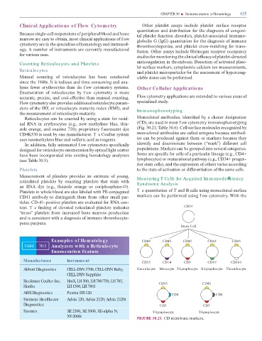
                        Reticulocytes can be counted by using a stain  or resid-                                               Monoclonal antibodies, identi  ed by a cluster designation

                   ual  RNA  in  erythrocytes  (e.g.,  new  methylene  blue,  thia-                                            (CD), are used in most f ow cytometry immunophenotyping

                   zole orange, and oxazine 750); proprietary f uorescent dye                                                  (Fig. 30.21;   able 30.6). Cell sur ace molecules recognized by

                   CD4K530 is used by one manu acturer. T e Coulter system                                                     monoclonal antibodies are called antigens because antibod-

                   uses neomethylene blue and sul uric acid as reagents.                                                       ies can be produced against them or markers because they

                        In addition,  ully automated f ow cytometers speci  cally                                              identi y  and  discriminate  between  (“mark”)  di  erent  cell

                   designed  or reticulocyte enumeration by optical light scatter                                              populations. Markers can be grouped into several categories.

                   have been incorporated into existing hematology analyzers                                                   Some are speci  c  or cells o  a particular lineage (e.g., CD4+

                   (see   able 30.5).                                                                                          lymphocytes) or maturational pathway (e.g., CD34+ progen-
                                                                                                                               itor stem cells), and the expression o  others varies according

                   Platelets                                                                                                   to the state o  activation or di  erentiation o  the same cells.


                   Measurement  o   platelets  provides  an  estimate  o   young,                                              Measuring T Cells for Acquired Im m unode  ciency

                   reticulated  platelets  by  counting  platelets  that  stain  with                                          Syndrom e Analysis

                   an  RNA  dye  (e.g.,  thiazole  orange  or  coriphosphine-O).

                   Platelets in whole blood are also labeled with PE-conjugated                                                T e quantitation o     and B cells using monoclonal sur ace

                   CD41 antibody to distinguish them  rom other small par-                                                     markers can be per ormed using f ow cytometry. With the

                   ticles. CD-41–positive platelets are evaluated  or RNA con-

                   tent. T e   nding o  elevated reticulated platelets indicates                                                                                          CD34

                   “stress”  platelets   rom  increased  bone  marrow  production

                   and is consistent with a diagnosis o  immune thrombocyto-

                   penic purpura.
                                                                                                                                                                        Stem Cell


                                                Examples of Hematology                                                              CD45               CD45              CD45                   CD45                  CD45


                       TABLE         30.5       Analyzers w ith a Reticulocyte

                                                Enumeration Feature


                       Manufacturer                            Instrument                                                           CD15               CD14               CD3                   CD19                  CD161


                       Abbott Diagnostics                      CELL-DYN 3700, CELL-DYN Ruby,                                   Granulocyte          Monocyte T-Lymphocyte B-Lymphocyte Thrombocyte

                                                               CELL-DYN Sapphire


                       Beckman Coulter Inc.                    HmX, LH 500, LH 700/750, LH 785,                                                    CD45                                    CD45

                       Horiba                                  LH 1500, LH 7801


                       ABX Diagnostics                         Pentra DX 120                                                                                   CD4                                      CD8

                       Siemens Healthcare                      Advia 120, Advia 2120, Advia 2120i

                       Diagnostics                                                                                                                  CD3                                     CD3


                       Sysmex                                  XE 2100, XE 5000, XE-alpha N,                                                  T-Lymphocyte                              T-Lymphocyte

                                                               XT-2000i                                                        FIGURE 30.21  CD membrane markers.
   646   647   648   649   650   651   652   653   654   655   656