
Page 655 - Clinical Hematology_ Theory _ Procedures ( PDFDrive )
P. 655
CHAPTER 30 ■ Instrumentation in Hematology 639
In 2000, CellaVision (Lund, Sweden) launched the
Di Master Octavia. T e system consists o an automated BOX 30.3
microscope with a 100× objective, a stepper motor and light
control unit, and a progressive three-chip CCD color cam-
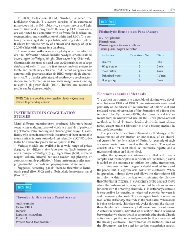
era connected to a computer with so ware or localization, Fibrinolytic Hemostasis Panel Assays
segmentation, and classi cation o white and RBCs. T e sys- α-2-Antiplasmin
tem processes eight slides per batch, utilizing a slide holder. Plasminogen
It allows or remote review o a smear and storage o up to Plasminogen activator inhibitor
20,000 slides with images in a database. issue plasminogen activator
In comparison with earlier attempts by other manu actur-
ers, the Di Master Octavia handles wedged smears stained Solution Container No. Time
according to the Wright, Wright-Giemsa, or May-Grünwald-
Giemsa staining protocols and uses ANNs trained on a large Fixative 2 30 s
database o cells. It was the rst image analysis system to Wright stain 3 3 min
locate and preclassi y cells into 15 di erent categories and Stain buffer 4 6 min
automatically precharacterize six RBC morphologic charac- Deionized water 5 1.5 min
teristics. T e platelet estimates and erythrocyte precharacter-
ization are per ormed in an overview image corresponding Drying stage 6 3 min
to eight high-power elds (100×). Review and release o
results can be done remotely.
Electromechanical Methods
NOTE: This is a good time to complete Review Questions T e earliest instruments to detect blood clotting were devel-
related to preceding content. oped between 1920 and 1940. T ese instruments were based
primarily on detection o the ormation o a brin clot and
replaced visual observation o the ormation o a brin clot
INSTRUMENTS IN COAGULATION in a test tube. By the mid-1960s, electromechanical instru-
STUDIES ments were in widespread use. In the 1970s, photo-optical
methods replaced electromechanical devices in most labora-
Many di erent manu acturers produced laboratory-based
coagulation analyzers, many o which are capable o per orm- tories, except student laboratories or as a backup method in
ing clottable, immunoassay, and chromogenic assays. T e di - routine laboratories.
culty with some instruments is that many o them are unable T e principle o electromechanical methodology is the
to transmit an industry-standard test identi er (LOINC code) measurement o conduction or impedance o an electri-
to the host laboratory in ormation system (LIS). cal current by the ormation o brin. An example o such
Various models are available in a wide range o prices a semiautomated instrument is the brometer. T is system
designed or di erent size laboratories. Each instrument consists o a 37°C heat block, an automatic pipette, and a
o ers unique advantages (e.g., high throughput, reduced mechanical mixer and timer block.
reagent volume, integral bar-code reader, cap piercing, or A er the appropriate containers are lled and plasma
automatic sample predilution). Many instruments o er user- samples and thromboplastin substrate are incubated, plasma
programmable methods and preprogrammed methods. is added to the substrate to initiate the timing mechanism.
Suggested screening panels include thrombotic hemo- T is timing mechanism triggers a digital readout time and
stasis panel (Box 30.2) and a brinolytic hemostasis panel the probe unit. T e probe arm holds two electrodes. When
(Box 30.3). in operation, it drops down and allows the electrodes to all
into place within the reaction well containing the plasma-
thromboplastin mixture. T e stationary probe does not move
when the instrument is in operation but unctions in con-
BOX 30.2 junction with the moving electrode. T is stationary electrode
is responsible or creating an electrical potential between it
Thrombotic Hemostasis Panel Assays and the moving electrode. T e moving electrode is located in
ront o the stationary electrode in the probe arm. When a test
Antithrombin is being per ormed, this electrode cycles through the plasma-
Factor VIII:C thromboplastin mixture every hal second until a clot orms.
Heparin A detection circuit is activated when a brin strand is ormed
Lupus anticoagulant between the two electrodes, thus completing the circuit. Circuit
Protein C activation stops the timer and prevents urther movement o
Protein S and ree protein S the moving electrode. Electromechanical methods, such as
the brometer, can be used or various coagulation assays.