Page 650 - Clinical Hematology_ Theory _ Procedures ( PDFDrive )
P. 650

634            PART 8  ■  Fundamentals of Hematological Analysis


               as a stain, using a similar method to that o  the  peroxidase                                               General Properties of Flow  Cytometry

               channel.  Lymphocytes,  which  have  been  labeled  by  the

               immunoperoxidase  reaction,  appear  between  the  unla-                                                    Most f ow-cell instruments can simultaneously analyze mul-

               beled lymphocyte population and the monocytes. Cells with                                                   tiple parameters at the rate o  5,000 to 10,000 cells/second.

               endogenous peroxidase such as neutrophils stain intensely                                                   T e cellular analysis yields quantitative data about the chem-

               and appear  ar to the right.                                                                                ical and physical properties o  individual cells, and a  er anal-
                                                                                                                           ysis, cells can be physically separated into subpopulations

                                                                                                                            or  urther study at the rate o  5,000 cells/second. T e major
               APPLICATIONS OF FLOW CYTOMETRY                                                                              advances in this technology are owing to several  actors:


               T e introduction o  the f ow cytometer into the clinical labo-                                              1.  T e ability to produce monoclonal antibodies resulted in

               ratory is a major technological advance. Flow cytometry is a                                                      the subsequent development o  speci  c sur ace markers

                eld that has evolved rapidly during the past three decades.                                                       or various subpopulations o  cells.

               Instruments based on the f ow cytometry principle were ini-                                                 2.  T e development o  new f uorescent probes  or DNA,

               tially designed to count and size cells. Later modi  cations                                                      RNA, and other cellular components increased the variety

               were designed to per orm di  erential leukocyte counts by                                                         o  possible applications at the molecular and cellular level.

               identi ying speci  c cytochemical reactions in the cells. T e                                               3.  T e expansion o  computer applications has improved the

               current  types  o   f ow  cytometry  instruments  can  analyze                                                    instrumentation technology, making it easier to operate

               cells  or many constituents and sort cells into subpopulations                                                    and more practical  or use in clinical as well as research

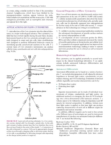
               (Fig. 30.20).                                                                                                     laboratories.


                                                                                                                           Hematological Applications


                                                                                                                           Flow cytometry can be applied practically to several tech-

                                                                                                                           niques in the clinical hematology laboratory. T ese appli-

                                                                                                                           cations  include  automated  leukocyte  di  erentiation  and

                                                                                                                           reticulocyte enumeration.


                                                                                                                           Autom ated Differentials


                                                                                                                           Automated di  erentials can be based on a variety o  princi-

                                                                                                                           ples. T ese include determination o  cell volume by electrical

                                                                                                                           impedance or  orward light scatter, cytochemistry or per-

                                                                                                                           oxidase staining, and VCS technology. Evaluation o  internal

                                                                                                                           cellular organelles and nuclear characteristics can be by:


                                                                                                                           ■    90-Degree laser scatter

                                                                                                                           ■    Polarizing laser light

                                                                                                                           ■    RF


                                                                                                                                Separate measurements can be made o  individual mea-

                                                                                                                           surements  o   volume,  conductivity,  and  light  scatter.  An

                                                                                                                           additional method is to integrate the three in VCS technol-

                                                                                                                           ogy into a three-dimensional (3D) leukocyte analysis. T e

                                                                                                                           volume aspect is by volumetric sizing by impedance and RF

                                                                                                                           opacity  or internal composition. In addition, helium-neon

                                                                                                                           laser light scatter is applied so that laser light can produce

                                                                                                                           scattering characteristics o  each cell at di  erent angles  or

                                                                                                                           granularity and nuclear structure.

                                                                                                                                In addition, di  erent reagents can be used to lyse certain

                                                                                                                           cells. Di  erent types o  technologies are used by instrument

                                                                                                                           manu acturers to produce an automated leukocyte di  eren-

                                                                                                                           tial. T ese include the  ollowing:

               FIGURE 30.20  Laser and cell-sorting schematic. In a f ow cytom-                                            Beckman Coulter: VCS

               etry system, stained cells f ow through a sample tube. As the cells

               and a stream o  saline solution leave the f ow chamber, they move                                           Abbott: MAPPS—0, 90, 10, and 90 degrees depolarized
               like a string o  beads in the center o  the sheath. T e f uorescence o                                      Roche (Sysmex): RF and DC

               the cells is detected by a sensor. Cells can be appropriately charged                                       Siemens  Healthcare  Diagnostics  ( ormerly  Bayer  and

               as they move through a charging collar or def ection plates. Sorting                                                echnicon):  peroxidase  staining;  optical  scatter  and
               o  the cells is accomplished by def ecting charged cells depending                                                 absorption;  basophils:  di  erential  lysis  laser  scatter

               on the charge (either positive or negative).                                                                       high and low
   645   646   647   648   649   650   651   652   653   654   655