
Page 674 - Clinical Hematology_ Theory _ Procedures ( PDFDrive )
P. 674
658 PART 8 ■ Fundamentals of Hematological Analysis
PCR assay has been run. DNA and other biomolecules can be
separated based on charge, size, and shape (see Figs. 31.3 and
31.4). DNA has a net negative charge and will migrate toward
the anode (positive pole). PCR products are loaded into an aga-
rose gel and electrophoresed. Ethidium bromide is a dye that
intercalates into nucleic acids and will f uoresce with an orange
color under ultraviolet (UV) irradiation. An image analyzer
uses UV light to capture computer images o the PCR products.
Melting curve analysis (MCA) is a method o assessing
the dissociation characteristics o double-stranded (DS)
DNA using a f uorophore during heating. When the tem-
perature is raised, the DS begins to dissociate, which leads
to a rise in the absorbance intensity, hyperchromicity. Factor
V Leiden coagulopathies are a good example, i a particular
mutation is known.
T e principle o MCA is that slightly mismatched DNA
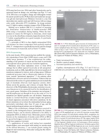
will denature at a lower temperature than per ectly matched FIGURE 31.3 PCR. Molecular gene structure o chromosomes 14
DNA. T e temperature is gradually increased, and the change and 18. Example o bcl-2 translocation detection by PCR. Lane 1, a
reactive lymphoid tissue showing no evidence o bcl-2 translocation
in f uorescence is measured, such as Factor V Leiden.
while in lanes 2 and 3 show 2 patients with ollicular lymphoma
DNA Sequencing having bcl-2 rearrangement. mw, molecular size markers. (From
McClatchey KD. Clinical Laboratory Medicine, 2nd ed, Philadelphia,
In the past decade, DNA sequencing has enabled the system- PA: Lippincott Williams & Wilkins, 2002.)
atic sequencing o more than 10,000 cancer exomes and 2,500
whole cancer genomes. T is has revolutionized the under- ■ Faster turnaround time
standing o the genetics o cancer and has lead to previously ■ Smaller required sample volumes
unrecognized cancer genes and new mutation signatures. ■ Increased speci city and sensitivity
Mutations arise rom replication errors or rom DNA
damage that is either repaired incorrectly or le unrepaired. Capillary electrophoresis (CE) (Figs. 31.5 and 31.6) is a
Leukemias, such as AML, typically have the lowest numbers o relatively new, power ul separation technique that is ideally
mutations compared to tumors, such as lung cancer. Di erent
mutational processes lead to idiosyncratic patterns o muta-
tions, termed “mutational signatures.” T ese patterns allow
identi cation o known and novel mutations. Features that can
characterize the action o a given mutation process include the
type o mutations observed, local sequence context, distri-
bution across the genome, and evidence o repair. Since the
discovery o the Philadelphia chromosome, mutation reoccur-
rence has proven to be a power ul tool or the identi cation o
new cancer genes. Estimates o the mutation rate in human
and B lymphocytes are thought to be on the order o 2 to 10
mutations per diploid genome per cell division. Sequencing
studies in normal blood has revealed insights into patterns
o clonal expansion associated with driver mutations. In the
blood, mutations ref ect about the relationship between aging,
particularly individuals older than 65 years o age, and the
typical epidemiological pattern seen in leukemias.
Many o the revolutionary changes that have occurred in
research in the biological sciences, particularly the Human FIGURE 31.4 PCR detection o actor V Leiden. Exon 10 o Factor
Genome Project, can be directly attributed to the ability to V gene is PCR ampli ed and cut with (D) resection enzyme MnlI.
manipulate DNA in de ned ways. Molecular genetic testing DNA rom normal (N) individuals contains MnlI, recognition site
ocuses on the examination o nucleic acids (DNA or RNA) (GAGG), which is lost in individuals carrying mutation. Undigested
by special techniques to determine whether a speci c nucle- DNA in normal (N) individuals shows 267 bp (upper bands) DNA
otide base sequence is present. ragment. A er resection enzyme treatment, normal individuals show
163 bp ragment (dashed arrow). In homozygous (Hom) patients,
T e applications o nucleic acid testing have expanded, digestion shows 200 bp ragment (solid arrow). In heterozygous (Het)
despite higher costs associated with testing, in various areas patients, both 163 bp and 200 bp ragments are seen. Internal control
o the clinical laboratory. T ese include genetic testing or o 67 bp in digested samples is seen in the bottom. U 5, undigested;
diagnosis and monitoring. Molecular testing has the ollow- D 5, digested. (From McClatchey KD. Clinical Laboratory Medicine,
ing advantages: 2nd ed, Philadelphia, PA: Lippincott Williams & Wilkins, 2002.)