Page 675 - Clinical Hematology_ Theory _ Procedures ( PDFDrive )
P. 675

CHAPTER 31  ■  Molecular Diagnostic Techniques and Applications                                      659


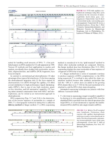
                                                                                                                                                                       FIGURE 31.5  PCR with capillary elec-

                                                                                                                                                                       trophoresis technique using three pairs
                                                                                                                                                                       o  primers  or the detection o  immu-

                                                                                                                                                                       noglobulin  heavy-chain  gene  rear-

                                                                                                                                                                       rangements. Frameworks I and II show

                                                                                                                                                                       a monoclonal peak, representing gene

                                                                                                                                                                       rearrangement. Framework III shows a
                                                                                                                                                                       polyclonal pattern. (From Sun   . Flow

                                                                                                                                                                       Cytometry, Immunohistochemistry, and

                                                                                                                                                                       Molecular  Genetics   or  Hematologic
                                                                                                                                                                       Neoplasms,  2nd  ed,  Philadelphia,  PA:

                                                                                                                                                                       Lippincott Williams & Wilkins, 2012.)


                   suited  or handling small amounts o  DNA. T e   rst pub-                                                    method is considered to be the “gold standard” method to

                   lished papers on DNA analysis by CE only appeared in 1988.                                                  which  other  molecular  methods  are  compared.  However,

                   Various CE methods and their applications to nucleic acid                                                   the Sanger method does have limitations (Box 31.3). DNA

                   analysis, speci  cally those dealing with nucleosides, nucleo-                                              sequencing displays the exact nucleotide or base sequence o

                   tides, oligonucleotides, and dsDNA (PCR)  ragments, have                                                    a  ragment o  DNA that is targeted.

                   been developed.                                                                                                  T e Sanger method uses a series o  enzymatic reactions

                        In contrast to conventional gel electrophoresis, CE takes                                              to produce segments o  DNA complementary to the DNA

                   advantage o  two types o  driving  orces: (1) the  orce causing                                             being sequenced. T is method is also called the chain ter-

                   the electrophoretic migration and (2) the  orce exerted by                                                  mination  method  because  a  er  synthetic  nucleotides  that

                   electroosmotic f ow (EOF) through the capillary. CE o  ers                                                  lack the −OH at 3′ carbon atom are added to the growing

                   several similarities to high-per ormance liquid chromatog-                                                  DNA strand, there is no 3′-OH  or the next nucleotide to be

                   raphy (HPLC), that is, ease o  use, high resolution, speed,                                                 attached to, and the DNA chain stops elongating.

                   on-line detection, and  ull automation capability. CE, hav-                                                      Automated sequencing techniques use primers with  our

                   ing taken essential components  rom both HPLC and elec-                                                     di  erent f uorescent labels.

                   trophoresis, can be viewed as an instrumental approach to

                   electrophoresis.                                                                                            1.  T e   rst step in sequencing a target is usually to ampli y it

                        DNA sequencing (Figs. 31.7 and 31.8) is the determina-                                                      by cloning or in vitro ampli  cation, usually PCR. Once the

                   tion o  the precise sequence o  nucleotides in a sample o                                                        ampli  ed DNA is puri  ed  rom the clinical specimen (the

                   DNA. T e most popular method  or doing this is called the                                                        target DNA), it is heat denatured to separate the double-

                   dideoxy method or Sanger method. T is DNA sequencing                                                             stranded DNA (dsDNA) into single strands (ssDNA).


                                                                                                                                                                        FIGURE 31.6  Splenic B-cell marginal

                                                                                                                                                                        zone  lymphoma.  Polymerase  chain

                                                                                                                                                                        reaction with capillary electrophoresis

                                                                                                                                                                        technique reveals a monoclonal peak in
                                                                                                                                                                        each o  the three  rameworks, represent-

                                                                                                                                                                        ing immunoglobulin heavy-chain gene

                                                                                                                                                                        rearrangement. (From  Sun   . Atlas  o

                                                                                                                                                                        Hematologic Neoplasms, New York, NY:
                                                                                                                                                                        Springer, 2009.)
   670   671   672   673   674   675   676   677   678   679   680