Page 678 - Clinical Hematology_ Theory _ Procedures ( PDFDrive )
P. 678

662            PART 8  ■  Fundamentals of Hematological Analysis


                                A
                                     gene A (normal)              CC TG AGG
                                                                                                                                BOX  31.4

                                                                     Ms t II site


                                     gene S (sickle)              CC TG T GG
                                                                                                                              Strengths and Limitations of FISH
                                                                 (no Ms t II site)

                                                                                                                              STRENGTHS OF FISH

                                B                   Restriction site                                                          ■   Is better than PCR  or deletions and inversions

                                                    absent in sickle-cell              β-globin                               ■   Is the next generation o  cytogenetic techniques a  er

                                                    β-globin                              gene
                                                                                                                                   banding
                                            Ms t II                   Ms t II Ms t II                                         ■   Can be correlated with morphology


                                                                                                                              ■   Can be used to assess DNA or mRNA

                                                                                                                              ■   Can be per ormed on metaphase spreads but can also
                                   gene A                 1.1kb                                                                    be done on para  n block sections


                                   gene S                   1.3kb
                                                                                                                              LIMITATIONS OF FISH

                                                                                                                              ■   Problems in tissue that has been decalci  ed or not   xed
                                C                                                                                                  in  ormalin

                                Southern blot                                                                                 ■   Requires a f uorescent microscope

                                of DNA cut with                                                                                   Probes must be speci  cally designed
                                Ms t II  and                                        β (1.3kb)                                 ■
                                                                                      S
                                hybridized                                                                                    ■   Can only be used to detect the presence or absence o
                                with β-globin                                       β (1.1kb)                                      previously identi  ed chromosomal aberrations
                                                                                      A
                                probe                                                                                         ■   Not good as PCR  or very small mutations

                                                                                                                              ■   Labor intensive

                                                                                                                              ■    issue that is   xed cannot be cultured  or metaphase
                                                                         Sickle-cell control
                                                                         Normal control                                            spreads

                                                                         Carrier
                                                                                                                              Modi  ed  rom Heriot K. Welcome to the beginning: molecular pathol-
                                                                         Affected individual                                  ogy  or the community hospital pathologist and medical technologist,


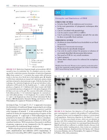
               FIGURE 31.9  Restriction  ragment length polymorphism (RFLP)                                                   ASCP Annual Meeting,   ampa, FL, 2014.
               caused by loss o  a restriction site. I  a mutation occurs in a cleav-

               age site  or a restriction enzyme, the pattern o  restriction  ragments

               di  ers  rom normal. A. T e mutation that causes sickle cell anemia
               results in the loss o  an MstII site in the β-globin gene. B. Samples

               o  DNA  rom individuals are treated with restriction endonucleases

               and then subjected to electrophoresis on gels. With the Southern blot

               technique, the restriction  ragments on the gel are hybridized with a

               radioactive cDNA probe  or the β-globin gene. T e sickle cell allele
               produces a  ragment o  1.3 kilobases (kb) when treated with MstII.

               A normal allele produces a  ragment o  1.1 kb (plus a  ragment o  0.2

               kb that is not seen on the gel). For a person with sickle cell disease,
               both alleles produce 1.3-kb restriction  ragments. In a normal per-

               son, both alleles produce 1.1-kb  ragments. For a carrier, both the

               1.3- and  1.1-kb  ragments are observed. (From Lieberman M, Ricer

               R (eds.). BRS Biochemistry, Molecular Biology, and Genetics, 6th ed,

               Philadelphia, PA: Lippincott Williams & Wilkins, 2013.)


               developed (Figs. 31.12 and 31.13). T is approach overcomes

               the limitations o  traditional Sanger sequencing by providing

               highly parallel sequencing with a separate sequence result  or

               every sequence o  interest. T is has positioned NGS as the                                                  FIGURE 31.10  Southern blot detection o    -cell receptor (  CR)

               method o  choice  or targeted resequencing o  regions o  the                                                gene rearrangement in a patient with peripheral   -cell lymphoma.

               human genome. T e approach o  NGS technologies has the                                                       wo separate lesions were investigated  or evidence o  a monoclonal

               potential to be more cost-e  ective and be able to simultane-                                                 -cell population. Restriction digestion in the BamHI (1, 2) and

               ously sequence complete genomes o  patients to deliver per-                                                 EcoRI (7, 8) show extra bands (arrow), which are not detected in

               sonalized medicine. NGS can produce thousands to millions                                                   the lane with DNA isolated  rom placenta (pl). Although HindIII
                                                                                                                           digest (4, 5) shows no extra band, two rearranged bands detected by
               o  genome sequences at one compared to the 96 sequences                                                     BamHI and EcoRI are su  cient to establish clonality. Because both

               processed by the traditional Sanger method.                                                                 lesions show similar banding pattern, these lesions are considered

                    NGS technologies permit analysis o  mutation, rearrange-                                               to be clonally identical. (Reprinted  rom McClatchey KD. Clinical

               ment,  ampli  cations  and  deletions  (DNA  sequencing),  or                                               Laboratory Medicine, 2nd ed, Philadelphia, PA: Lippincott Williams

               coding and noncoding RNA (RNA sequencing). Most clini-                                                      & Wilkins, 2002, with permission.)
   673   674   675   676   677   678   679   680   681   682   683