
Page 252 - Review of Medical Microbiology and Immunology ( PDFDrive )
P. 252
mebooksfree.com
mebooksfree.com
mebooksfree.com
mebooksfree.com
mebooksfree.com
mebooksfree.com
mebooksfree.com mebooksfree.com mebooksfree.com However, the prophage is not permanently integrated. It 241 mebooksfree.com
mebooksfree.com
mebooksfree.com
CHAPTER 29 Replication
β-Phage
β-Phage
can be induced to resume its replicative cycle by the action
genes
genes
Diphtheria
of ultraviolet (UV) light and certain chemicals that damage
Diphtheria
toxin
toxin gene
DNA. UV light induces the synthesis of a protease, which
cleaves the repressor. Early genes then function, including
from the cell DNA. The virus then completes its replicative
cycle, leading to the production of progeny virus and lysis
Bacterial Lysogenic conversion the genes coding for the enzymes that excise the prophage
of the cell.
chromosome Diphtheria toxin Relationship of Lysogeny in Bacteria to
mebooksfree.com
mebooksfree.com mebooksfree.com mebooksfree.com Latency in Human Cells mebooksfree.com mebooksfree.com
β phage carrying
genes integrated
diphtheria toxin gene
into chromosome
infects C. diphtheriae
of C. diphtheriae;
not lysogenized by
Members of the herpesvirus family, such as herpes simplex
bacterium becomes
β phage; bacterium is
pathogenic.
nonpathogenic prior
virus (HSV), varicella-zoster virus, cytomegalovirus
to infection by β phage.
(CMV), and Epstein–Barr virus, exhibit latency—the phe-
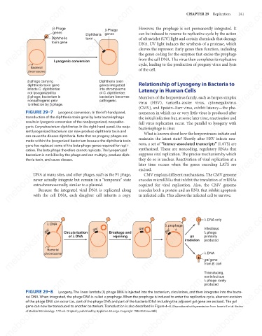
FIGURE 29–7
nomenon in which no or very little virus is produced after
Lysogenic conversion. In the left-hand panel,
the initial infection but, at some later time, reactivation and
transduction of the diphtheria toxin gene by beta bacteriophage
results in lysogenic conversion of the nonlysogenized, nonpatho-
genic Corynebacterium diphtheriae. In the right-hand panel, the recip-
bacteriophage is clear.
ient lysogenized bacterium can now produce diphtheria toxin and
What is known about how the herpesviruses initiate and
can cause the disease diphtheria. Note that no progeny phages are full virus replication occur. The parallel to lysogeny with
maintain the latent state? Shortly after HSV infects neu-
made within the lysogenized bacterium because the diphtheria toxin
mebooksfree.com
mebooksfree.com
mebooksfree.com mebooksfree.com mebooksfree.com they do so is unclear. Reactivation of viral replication at a mebooksfree.com
rons, a set of “latency-associated transcripts” (LATS) are
gene has replaced some of the beta-phage genes required for repli-
synthesized. These are noncoding, regulatory RNAs that
cation. The beta phage therefore cannot replicate. The lysogenized
suppress viral replication. The precise mechanism by which
bacterium is not killed by the phage and can multiply, produce diph-
theria toxin, and cause disease.
later time occurs when the genes encoding LATS are
excised.
CMV employs different mechanisms. The CMV genome
DNA at many sites, and other phages, such as the P1 phage,
never actually integrate but remain in a “temperate” state
encodes microRNAs that inhibit the translation of mRNAs
extrachromosomally, similar to a plasmid.
required for viral replication. Also, the CMV genome
Because the integrated viral DNA is replicated along
encodes both a protein and an RNA that inhibit apoptosis
in infected cells. This allows the infected cell to survive.
with the cell DNA, each daughter cell inherits a copy.
mebooksfree.com mebooksfree.com Circularization gal Breakage and gal mebooksfree.com λ DNA only mebooksfree.com
mebooksfree.com
mebooksfree.com
λ DNA
λ prophage
Infectious
gal
gal
λ phage
of k DNA
rejoining
primarily
UV
produced
irradiation
Bacterial
λ DNA
chromosome
gal gene
from E. coli
Transducing,
noninfectious
mebooksfree.com mebooksfree.com mebooksfree.com mebooksfree.com mebooksfree.com mebooksfree.com
λ phage rarely
produced
FIGURE 29–8
Lysogeny. The linear lambda (λ) phage DNA is injected into the bacterium, circularizes, and then integrates into the bacte-
rial DNA. When integrated, the phage DNA is called a prophage. When the prophage is induced to enter the replicative cycle, aberrant excision
of the phage DNA can occur (i.e., part of the phage DNA and part of the bacterial DNA including the adjacent gal gene are excised). The gal
gene can now be transduced to another bacterium. Transduction is also described in Figure 4–4. (Reproduced with permission from Jawetz E et al. Review
of Medical Microbiology. 17th ed. Originally published by Appleton & Lange. Copyright 1986 McGraw-Hill.)
mebooksfree.com mebooksfree.com mebooksfree.com mebooksfree.com mebooksfree.com mebooksfree.com