


Page 107 - Textbook of Pathology, 6th Edition
P. 107
91  CHAPTER 4


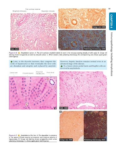
           Figure 4.16  Amyloidosis spleen. A, The pink acellular amyloid material is seen in the red pulp causing atrophy of while pulp. B, Congo red
           staining shows Congophilia as seen by red-pink colour. C, When viewed under polarising microscopy the corresponding area shows apple-green  Immunopathology Including Amyloidosis
           birefringence.

               Later, as the deposits increases, they compress the  However, hepatic function remains normal even at an
            cords of hepatocytes so that eventually the liver cells  advanced stage of the disease.
            are shrunken and atrophic and replaced by amyloid.      To a lesser extent, portal tracts and Kupffer cells are
                                                                 involved in amyloidosis.


           Figure 4.17  Amyloidosis of the liver. A, The deposition is extensive
           in the space of Disse causing compression and pressure atrophy of
           hepatocytes. B, Congo red staining shows congophilia which under
           polarising microscopy. C, shows apple-green birefringence.

