


Page 111 - Textbook of Pathology, 6th Edition
P. 111
Normal Fluid Pressures                                 There is considerable pressure gradient at the two ends of  95
           1. OSMOTIC PRESSURE. This is the pressure exerted by  capillary loop—being higher at the arteriolar end (average
           the chemical constituents of the body fluids. Accordingly,  32 mmHg) than at the venular end (average 12 mmHg).
           osmotic pressure may be of the following types (Fig. 5.3,A):  Tissue tension is the hydrostatic pressure of interstitial
              Crystalloid osmotic pressure exerted by electrolytes  fluid and is lower than the hydrostatic pressure in the
           present in the ECF and comprises the major portion of the  capillary at either end (average 4 mmHg).       CHAPTER 5
           total osmotic pressure.                                 Effective hydrostatic pressure is the difference between
              Colloid osmotic pressure (Oncotic pressure) exerted by  the higher hydrostatic pressure in the capillary and the lower
           proteins present in the ECF and constitutes a small part of  tissue tension; it is the force that drives fluid through the capillary
           the total osmotic pressure but is more significant  wall into the interstitial space.
           physiologically. Since the protein content of the plasma is
           higher than that of interstitial fluid, oncotic pressure of  Normal Fluid Exchanges
           plasma is higher (average 25 mmHg) than that of interstitial  Normally, the fluid exchanges between the body
           fluid (average 8 mmHg).                             compartments take place as under:
              Effective oncotic pressure is the difference between the  At the arteriolar end of the capillary, the balance between
           higher oncotic pressure of plasma and the lower oncotic  the hydrostatic pressure (32 mmHg) and plasma oncotic
           pressure of interstitial fluid and is the force that tends to draw  pressure (25 mmHg) is the hydrostatic pressure of 7 mmHg
           fluid into the vessels.
                                                               which is the outward-driving force so that a small quantity
           2. HYDROSTATIC PRESSURE. This is the capillary blood  of fluid and solutes leave the vessel to enter the interstitial
           pressure.                                           space.                                                 Derangements of Homeostasis and Haemodynamics


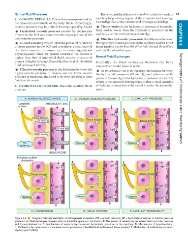
           Figure 5.3  Diagrammatic representation of pathogenesis of oedema (OP = oncotic pressure; HP = hydrostatic pressure). A, Normal pressure
           gradients and fluid exchanges between plasma, interstitial space and lymphatics. B, Mechanism of oedema by decreased plasma oncotic pressure
           and hypoproteinaemia. C, Mechanism of oedema by increased hydrostatic pressure in the capillary. D, Mechanism of lymphoedema.
           E, Mechanism by tissue factors (increased oncotic pressure of interstitial fluid and lowered tissue tension). F, Mechanism of oedema by increased
           capillary permeability.

