


Page 112 - Textbook of Pathology, 6th Edition
P. 112
96 At the venular end of the capillary, the balance between the produces a depression known as pitting oedema. The other
hydrostatic pressure (12 mmHg) and plasma oncotic pressure variety is non-pitting or solid oedema in which no pitting is
(25 mmHg) is the oncotic pressure of 13 mmHg which is the produced on pressure e.g. in myxoedema, elephantiasis.
inward-driving force so that the fluid and solutes re-enter The oedema may be of 2 main types:
the plasma. 1. Localised when limited to an organ or limb e.g. lymphatic
The tissue fluid left after exchanges across the capillary oedema, inflammatory oedema, allergic oedema.
wall escapes into the lymphatics from where it is finally 2. Generalised (anasarca or dropsy) when it is systemic in
drained into venous circulation. distribution, particularly noticeable in the subcutaneous
tissues e.g. renal oedema, cardiac oedema, nutritional
Tissue factors i.e. oncotic pressure of interstitial fluid and
SECTION I
tissue tension, are normally small and insignificant forces oedema.
Besides, there are a few special forms of oedema (e.g.
opposing the plasma hydrostatic pressure and capillary pulmonary oedema, cerebal oedema) discussed later.
hydrostatic pressure, respectively.
Depending upon fluid composition, oedema fluid may
be:
DISTURBANCES OF BODY FLUIDS transudate which is more often the case, such as in oedema
of cardiac and renal disease; or
The common derangements of body fluid are as follows: exudate such as in inflammatory oedema.
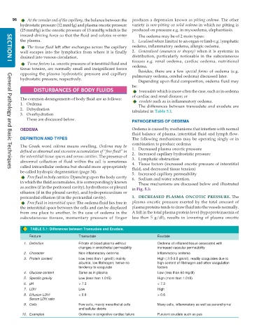
1. Oedema The differences between transudate and exudate are
2. Dehydration tabulated in Table 5.1.
3. Overhydration
These are discussed below.
PATHOGENESIS OF OEDEMA
OEDEMA Oedema is caused by mechanisms that interfere with normal
fluid balance of plasma, interstitial fluid and lymph flow.
DEFINITION AND TYPES The following mechanisms may be operating singly or in
combination to produce oedema:
The Greek word oidema means swelling. Oedema may be 1. Decreased plasma oncotic pressure
defined as abnormal and excessive accumulation of “free fluid” in 2. Increased capillary hydrostatic pressure
the interstitial tissue spaces and serous cavities. The presence of 3. Lymphatic obstruction
abnormal collection of fluid within the cell is sometimes 4. Tissue factors (increased oncotic pressure of interstitial
called intracellular oedema but should more appropriately fluid, and decreased tissue tension)
General Pathology and Basic Techniques
be called hydropic degeneration (page 34). 5. Increased capillary permeability
Free fluid in body cavities: Dpending upon the body cavity 6. Sodium and water retention.
in which the fluid accumulates, it is correspondingly known These mechanisms are discussed below and illustrated
as ascites (if in the peritoneal cavity), hydrothorax or pleural in Fig. 5.3:
effusion (if in the pleural cavity), and hydropericardium or
pericardial effusion (if in the pericardial cavity). 1. DECREASED PLASMA ONCOTIC PRESSURE. The
Free fluid in interstitial space: The oedema fluid lies free in plasma oncotic pressure exerted by the total amount of
the interstitial space between the cells and can be displaced plasma proteins tends to draw fluid into the vessels normally.
from one place to another. In the case of oedema in the A fall in the total plasma protein level (hypoproteinaemia of
subcutaneous tissues, momentary pressure of finger less than 5 g/dl), results in lowering of plasma oncotic
TABLE 5.1: Differences between Transudate and Exudate.
Feature Transudate Exudate
1. Definition Filtrate of blood plasma without Oedema of inflamed tissue associated with
changes in endothelial permeability increased vascular permeability
2. Character Non-inflammatory oedema Inflammatory oedema
3. Protein content Low (less than 1 gm/dl); mainly High ( 2.5-3.5 gm/dl), readily coagulates due to
albumin, low fibrinogen; hence no high content of fibrinogen and other coagulation
tendency to coagulate factors
4. Glucose content Same as in plasma Low (less than 60 mg/dl)
5. Specific gravity Low (less than 1.015) High (more than 1.018)
6. pH > 7.3 < 7.3
7. LDH Low High
8. Effusion LDH/ < 0.6 > 0.6
Serum LDH ratio
9. Cells Few cells, mainly mesothelial cells Many cells, inflammatory as well as parenchymal
and cellular debris
10. Examples Oedema in congestive cardiac failure Purulent exudate such as pus

