


Page 115 - Textbook of Pathology, 6th Edition
P. 115
iv) Decreased capillary hydrostatic pressure associated with but is largely due to excessive reabsorption of sodium and 99
increased renal vascular resistance. water in the renal tubules via renin-angiotensin-aldosterone
The examples of oedema by these mechanims are as mechanism. The protein content of oedema fluid in
under: glomerulonephritis is quite low (less than 0.5 g/dl).
i) Oedema of cardiac disease e.g. in congestive cardiac failure. The nephritic oedema is usually mild as compared to
ii) Ascites of liver disease e.g. in cirrhosis of liver. nephrotic oedema and begins in the loose tissues such as on
iii) Oedema of renal disease e.g. in nephrotic syndrome, acute the face around eyes, ankles and genitalia. Oedema in these CHAPTER 5
glomerulonephritis. conditions is usually not affected by gravity (unlike cardiac
oedema).
PATHOGENESIS AND MORPHOLOGY OF The salient differences between the nephrotic and
IMPORTANT TYPES OF OEDEMA
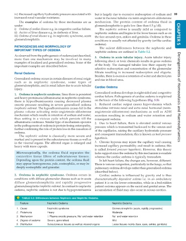
nephritic oedema are outlined in Table 5.2.
As observed from the pathogenesis of oedema just described, 3. Oedema in acute tubular injury. Acute tubular injury
more than one mechanism may be involved in many
examples of localised and generalised oedema. Some of the following shock or toxic chemicals results in gross oedema
important examples are described below. of the body. The damaged tubules lose their capacity for
selective reabsorption and concentration of the glomerular
Renal Oedema filtrate resulting in increased reabsorption and oliguria.
Besides, there is excessive retention of water and electrolytes
Generalised oedema occurs in certain diseases of renal origin and rise in blood urea.
such as in nephrotic syndrome, some types of
glomerulonephritis, and in renal failure due to acute tubular Cardiac Oedema
injury.
Generalised oedema develops in right-sided and congestive
1. Oedema in nephrotic syndrome. Since there is persistent
and heavy proteinuria (albuminuria) in nephrotic syndrome, cardiac failure. Pathogenesis of cardiac oedema is explained Derangements of Homeostasis and Haemodynamics
there is hypoalbuminaemia causing decreased plasma on the basis of the following hypotheses (Fig. 5.5):
oncotic pressure resulting in severe generalised oedema 1. Reduced cardiac output causes hypovolaemia which
(nephrotic oedema). The hypoalbuminaemia causes fall in the stimulates intrinsic-renal and extra-renal hormonal (renin-
plasma volume activating renin-angiotensin-aldosterone angiotensin-aldosterone) mechanisms as well as ADH
mechanism which results in retention of sodium and water, secretion resulting in sodium and water retention and
thus setting in a vicious cycle which persists till the consequent oedema.
albuminuria continues. Similar type of mechanism operates 2. Due to heart failure, there is elevated central venous
in the pathogenesis of oedema in protein-losing enteropathy, pressure which is transmitted backward to the venous end
further confirming the role of protein loss in the causation of of the capillaries, raising the capillary hydrostatic pressure
oedema.
The nephrotic oedema is classically more severe and and consequent transudation; this is known as back pressure
marked and is present in the subcutaneous tissues as well as hypothesis.
in the visceral organs. The affected organ is enlarged and 3. Chronic hypoxia may injure the capillary wall causing
heavy with tense capsule. increased capillary permeability and result in oedema; this
is called forward pressure hypothesis. However, this theory
Microscopically, the oedema fluid separates the lacks support since the oedema by this mechanism is exudate
connective tissue fibres of subcutaneous tissues. whereas the cardiac oedema is typically transudate.
Depending upon the protein content, the oedema fluid In left heart failure, the changes are, however, different.
may appear homogeneous, pale, eosinophilic, or may be There is venous congestion, particularly in the lungs, so that
deeply eosinophilic and granular. pulmonary oedema develops rather than generalised oedema
(described below).
2. Oedema in nephritic syndrome. Oedema occurs in Cardiac oedema is influenced by gravity and is thus
conditions with diffuse glomerular disease such as in acute characteristically dependent oedema i.e. in an ambulatory
diffuse glomerulonephritis and rapidly progressive patient it is on the lower extremities, while in a bed-ridden
glomerulonephritis (nephritic oedema). In contrast to nephrotic patient oedema appears on the sacral and genital areas. The
oedema, nephritic oedema is not due to hypoproteinaemia accumulation of fluid may also occur in serous cavities.
TABLE 5.2: Differences between Nephrotic and Nephritic Oedema.
Feature Nephrotic Oedema Nephritic Oedema
1. Cause Nephrotic syndrome Glomerulonephritis (acute, rapidly progressive)
2. Proteinuria Heavy Moderate
3. Mechanism ↓ Plasma oncotic pressure, Na and water retention Na and water retention
+
+
4. Degree of oedema Severe, generalised Mild
5. Distribution Subcutaneous tissues as well as visceral organs Loose tissues mainly (face, eyes, ankles, genitalia)

