


Page 117 - Textbook of Pathology, 6th Edition
P. 117
101


                                                                                                                      CHAPTER 5


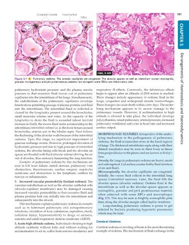
           Figure 5.7  Pulmonary oedema. The alveolar capillaries are congested. The alveolar spaces as well as interstitium contain eosinophilic,
           granular, homogeneous and pink proteinaceous oedema fluid alongwith some RBCs and inflammatory cells.


           pulmonary hydrostatic pressure and the plasma oncotic  respiratory ill-effects. Commonly, the deleterious effects
           pressure so that excessive fluid moves out of pulmonary  begin to appear after an altitude of 2500 metres is reached.
           capillaries into the interstitium of the lungs. Simultaneously,  These changes include appearance of oedema fluid in the  Derangements of Homeostasis and Haemodynamics
           the endothelium of the pulmonary capillaries develops  lungs, congestion and widespread minute haemorrhages.
           fenestrations permitting passage of plasma proteins and fluid  These changes can cause death within a few days. The under-
           into the interstitium. The interstitial fluid so collected is  lying mechanism appears to be anoxic damage to the
           cleared by the lymphatics present around the bronchioles,  pulmonary vessels. However, if acclimatisation to high
           small muscular arteries and veins. As the capacity of the  altitude is allowed to take place, the individual develops
           lymphatics to drain the fluid is exceeded (about ten-fold  polycythaemia, raised pulmonary arterial pressure, increased
           increase in fluid), the excess fluid starts accumulating in the  pulmonary ventilation and a rise in heart rate and increased
           interstitium (interstitial oedema) i.e. in the loose tissues around  cardiac output.
           bronchioles, arteries and in the lobular septa. Next follows
           the thickening of the alveolar walls because of the interstitial  MORPHOLOGIC FEATURES. Irrespective of the under-
           oedema. Upto this stage, no significant impairment of  lying mechanism in the pathogenesis of pulmonary
           gaseous exchange occurs. However, prolonged elevation of  oedema, the fluid accumulates more in the basal regions
           hydrostatic pressure and due to high pressure of interstitial  of lungs. The thickened interlobular septa along with their
           oedema, the alveolar lining cells break and the alveolar air  dilated lymphatics may be seen in chest X-ray as linear
           spaces are flooded with fluid (alveolar oedema) driving the air  lines perpendicular to the pleura and are known as Kerley’s
           out of alveolus, thus seriously hampering the lung function.  lines.
              Examples of pulmonary oedema by this mechanism are  Grossly, the lungs in pulmonary oedema are heavy, moist
           seen in left heart failure, mitral stenosis, pulmonary vein  and subcrepitant. Cut surface exudes frothy fluid (mixture
           obstruction, thyrotoxicosis, cardiac surgery, nephrotic  of air and fluid).
           syndrome and obstruction to the lymphatic outflow by  Microscopically, the alveolar capillaries are congested.
           tumour or inflammation.                               Initially, the excess fluid collects in the interstitial lung
                                                                 spaces (interstitial oedema). Later, the fluid fills the
           2. Increased vascular permeability (Irritant oedema). The  alveolar spaces (alveolar oedema). Oedema fluid in the
           vascular endothelium as well as the alveolar epithelial cells  interstitium as well as the alveolar spaces appears as
           (alveolo-capillary membrane) may be damaged causing   eosinophilic, granular and pink proteinaceous material,
           increased vascular permeability so that excessive fluid and  often admixed with some RBCs and macrophages
           plasma proteins leak out, initially into the interstitium and  (Fig. 5.7). This may be seen as brightly eosinophilic pink
           subsequently into the alveoli.                        lines along the alveolar margin called hyaline membrane.
              This mechanism explains pulmonary oedema in examples  Long-standing pulmonary oedema is prone to get
           such as in fulminant pulmonary and extrapulmonary     infected by bacteria producing hypostatic pneumonia
           infections, inhalation of toxic substances, aspiration, shock,
           radiation injury, hypersensitivity to drugs or antisera,  which may be fatal.
           uraemia and adult respiratory distress syndrome (ARDS).
                                                               Cerebral Oedema
           3. Acute high altitude oedema. Individuals climbing to high
           altitude suddenly without halts and without waiting for  Cerebral oedema or swelling of brain is the most threatening
           acclimatisation to set in, suffer from serious circulatory and  example of oedema. The mechanism of fluid exchange in the

