


Page 120 - Textbook of Pathology, 6th Edition
P. 120
104     serum concentration of bicarbonate; and             Clinically, there is peripheral vasodilatation and raised
              partial pressure of CO  that determines the concentration  intracranial pressure. If there is severe CO  retention, patients
                                                                                                  2
                               2
           of carbonic acid.                                   may develop confusion, drowsiness and coma. The arterial
              Accordingly, the disorders of the pH of the blood, termed  Pco  level is raised.
                                                                  2
           as acidosis (blood pH below 7.4) and alkalosis (blood pH
           above 7.4), can be of 2 types:                      Respiratory Alkalosis
           1. Alterations in the blood bicarbonate levels: These are  A rise in the blood pH occurring due to lowered Pco
           metabolic acidosis and alkalosis.                   consequent to hyperventilation of the lungs (excess removal 2
           2. Alteration in Pco  (which depends upon the ventilatory  of CO ) is called respiratory alkalosis. This is encountered in
     SECTION I
                                                                    2
                           2
           function of the lungs): These are respiratory acidosis and  the following conditions:
           alkalosis.                                              Hysterical overbreathing
              Thus, abnormalities in acid-base homeostasis produce  Working at high temperature
           following 4 principal metabolic syndromes which have    At high altitude
           diverse clinical manifestations due to pathophysiologic  Meningitis, encephalitis
           derangements:                                           Salicylate intoxication
                                                                  Clinically, the patients with respiratory alkalosis are
           Metabolic Acidosis                                  characterised by peripheral vasoconstriction and consequent
                                                               pallor, lightheadedness and tetany. The arterial Pco  is
           A fall in the blood pH due to metabolic component is brought  lowered.                              2
                                                      +
           about by fall of bicarbonate level and excess of H  ions in
           the blood. This occurs in the following situations:      HAEMODYNAMIC DERANGEMENTS
              Production of large amounts of lactic acid (lactic acido-
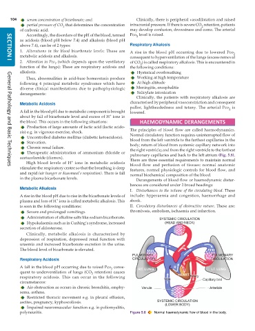
           sis) e.g. in vigorous exercise, shock.              The principles of blood flow are called haemodynamics.
              Uncontrolled diabetes mellitus (diabetic ketoacidosis).  Normal circulatory function requires uninterrupted flow of
              Starvation.                                      blood from the left ventricle to the farthest capillaries in the
                                                               body; return of blood from systemic capillary network into
              Chronic renal failure.                           the right ventricle; and from the right ventricle to the farthest
              Therapeutic administration of ammonium chloride or
           acetazolamide (diamox).                             pulmonary capillaries and back to the left atrium (Fig. 5.8).
                                                               There are three essential requirements to maintain normal
                                   +
              High blood levels of H  ions in metabolic acidosis  blood flow and perfusion of tissues: normal anatomic
           stimulate the respiratory centre so that the breathing is deep  features, normal physiologic controls for blood flow, and
     General Pathology and Basic Techniques
           and rapid (air hunger or Kussmaul’s respiration). There is fall  normal biochemical composition of the blood.
           in the plasma bicarbonate levels.                      Derangements of blood flow or haemodynamic distur-
                                                               bances are considered under 2 broad headings:
           Metabolic Alkalosis
                                                               I. Disturbances in the volume of the circulating blood. These
           A rise in the blood pH due to rise in the bicarbonate levels of  include: hyperaemia and congestion, haemorrhage and
                            +
           plasma and loss of H  ions is called metabolic alkalosis. This  shock.
           is seen in the following conditions:                II. Circulatory disturbances of obstructive nature. These are:
              Severe and prolonged vomitings.                  thrombosis, embolism, ischaemia and infarction.
              Administration of alkaline salts like sodium bicarbonate.
              Hypokalaemia such as in Cushing’s syndrome, increased
           secretion of aldosterone.
              Clinically, metabolic alkalosis is characterised by
           depression of respiration, depressed renal function with
           uraemia and increased bicarbonate excretion in the urine.
           The blood level of bicarbonate is elevated.

           Respiratory Acidosis
           A fall in the blood pH occurring due to raised Pco  conse-
                                                      2
           quent to underventilation of lungs (CO  retention) causes
                                             2
           respiratory acidosis. This can occur in the following
           circumstances:
              Air obstruction as occurs in chronic bronchitis, emphy-
           sema, asthma.
              Restricted thoracic movement e.g. in pleural effusion,
           ascites, pregnancy, kyphoscoliosis.
              Impaired neuromuscular function e.g. in poliomyelitis,
           polyneuritis.                                       Figure 5.8  Normal haemodynamic flow of blood in the body.

