


Page 204 - Textbook of Pathology, 6th Edition
P. 204
188   action of the trophozoite and have necrotic bed. Such
            chronic amoebic ulcers are described as flask-shaped
            ulcers due to their shape. The margin of the ulcer shows
            inflammatory response consisting of admixture of poly-
            morphonuclear as well as mononuclear cells besides the
            presence of trophozoites of Entamoeba histolytica (Fig. 7.10).
               Amoeboma is the inflammatory thickening of the wall
            of large bowel resembling carcinoma of the colon.
            Microscopically, the lesion consists of inflammatory
     SECTION I
            granulation tissue, fibrosis and clusters of trophozoites at
            the margin of necrotic with viable tissue.
               Amoebic liver abscess may be formed by invasion of
            the radicle of the portal vein by trophozoites. Amoebic
            liver abscess may be single or multiple (Chapter 21). The
            amoebic abscess contains yellowish-grey amorphous
            liquid material in which trophozoites are identified at the
            junction of the viable and necrotic tissue.
               Other sites where spread of amoebic infection may  Figure 7.10  Amoebic colitis. Section from margin of amoebic ulcer
            occur are peritonitis by perforation of amoebic ulcer of  shows necrotic debris, acute inflammatory infiltrate and a few trophozoites
            colon, extension to the lungs and pleura by rupture of  of Entamoeba histolytica (arrow).
            amoebic liver abscess, haematogenous spread to cause  malariae. While  Plasmodium falciparum causes malignant
            amoebic carditis and cerebral lesions, cutaneous   malaria, the other three species produce benign form of
            amoebiasis via spread of rectal amoebiasis or from anal  illness. These parasites are transmitted by bite of female
            intercourse.                                       Anopheles mosquito. The disease is endemic in several parts
                                                               of the world, especially in tropical Africa, parts of South and
           MALARIA
                                                               Central America, India and South-East Asia.
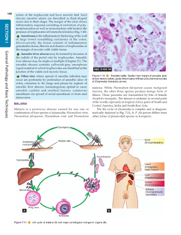
           Malaria is a protozoal disease caused by any one or    The life cycle of plasmodia is complex and is diagram-
           combination of four species of plasmodia: Plasmodium vivax,  matically depicted in Fig. 7.11, A. P. falciparum differs from
           Plasmodium falciparum, Plasmodium ovale and  Plasmodium  other forms of plasmodial species in 4 respects:
     General Pathology and Basic Techniques


           Figure 7.11  Life cycle of malaria (A) and major pathological changes in organs (B).

