


Page 206 - Textbook of Pathology, 6th Edition
P. 206
190                                                      are ingested by pigs or they infect vegetables. These eggs
                                                               then develop into larval stages in the host, spread by blood
                                                               to any site in the body and form cystic larvae termed
                                                               cysticercus cellulosae. Human beings may acquire infection
                                                               by the larval stage by eating undercooked pork (‘measly
                                                               pork’), by ingesting uncooked contaminated vegetables, and
                                                               sometimes, by autoinfection.


                                                                 MORPHOLOGIC FEATURES. The cysticercus may be
     SECTION I
                                                                 single or there may be multiple cysticerci in the different
                                                                 tissues of the body. The cysts may occur virtually
                                                                 anywhere in body and accordingly produce symptoms;
                                                                 most common sites are the brain, skeletal muscle and skin.
                                                                 Cysticercus consists of a round to oval white cyst, about
                                                                 1 cm in diameter, contains milky fluid and invaginated
                                                                 scolex with birefringent hooklets. The cysticercus may
                                                                 remain viable for a long time and incite no inflammation.
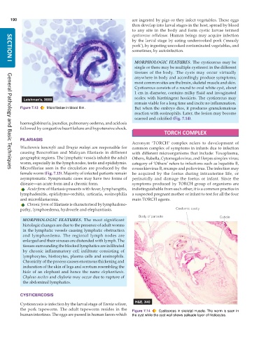
           Figure 7.13  Microfilariae in blood film.             But when the embryo dies, it produces granulomatous
                                                                 reaction with eosinophils. Later, the lesion may become
                                                                 scarred and calcified (Fig. 7.14).
           haemoglobinuria, jaundice, pulmonary oedema, and acidosis
           followed by congestive heart failure and hypotensive shock.
                                                                               TORCH COMPLEX
           FILARIASIS
                                                               Acronym ‘TORCH’ complex refers to development of
           Wuchereria bancrofti and  Brugia malayi are responsible for  common complex of symptoms in infants due to infection
           causing Bancroftian and Malayan filariasis in different  with different microorganisms that include: Toxoplasma,
           geographic regions. The lymphatic vessels inhabit the adult  Others, Rubella, Cytomegalovirus, and Herpes simplex virus;
           worm, especially in the lymph nodes, testis and epididymis.  category of ‘Others’ refers to infections such as hepatitis B,
           Microfilariae seen in the circulation are produced by the  coxsackievirus B, mumps and poliovirus. The infection may
     General Pathology and Basic Techniques
           female worm (Fig. 7.13). Majority of infected patients remain  be acquired by the foetus during intrauterine life, or
           asymptomatic. Symptomatic cases may have two forms of  perinatally and damage the foetus or infant. Since the
           disease—an acute form and a chronic form.           symptoms produced by TORCH group of organisms are
              Acute form of filariasis presents with fever, lymphangitis,  indistinguishable from each other, it is a common practice in
           lymphadenitis, epididymo-orchitis, urticaria, eosinophilia  a suspected pregnant mother or infant to test for all the four
           and microfilariaemia.                               main TORCH agents.
              Chronic form of filariasis is characterised by lymphadeno-
           pathy, lymphoedema, hydrocele and elephantiasis.

            MORPHOLOGIC FEATURES. The most significant
            histologic changes are due to the presence of adult worms
            in the lymphatic vessels causing lymphatic obstruction
            and lymphoedema. The regional lymph nodes are
            enlarged and their sinuses are distended with lymph. The
            tissues surrounding the blocked lymphatics are infiltrated
            by chronic inflammatory cell infiltrate consisting of
            lymphocytes, histiocytes, plasma cells and eosinophils.
            Chronicity of the process causes enormous thickening and
            induration of the skin of legs and scrotum resembling the
            hide of an elephant and hence the name  elephantiasis.
            Chylous ascites and chyluria may occur due to rupture of
            the abdominal lymphatics.


           CYSTICERCOSIS

           Cysticercosis is infection by the larval stage of Taenia solium,
           the pork tapeworm. The adult tapeworm resides in the  Figure 7.14  Cysticercus in skeletal muscle. The worm is seen in
           human intestines. The eggs are passed in human faeces which  the cyst while the cyst wall shows palisade layer of histiocytes.

