


Page 263 - Textbook of Pathology, 6th Edition
P. 263
247


                                                                                                                     CHAPTER 9


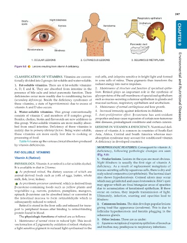
           Figure 9.8  Lesions resulting from vitamin A deficiency.

           CLASSIFICATION OF VITAMINS. Vitamins are conven-    rod cells, and iodopsins sensitive in bright light and formed
           tionally divided into 2 groups: fat-soluble and water-soluble.  in cone cells of retina. These pigments then transform the
           1. Fat-soluble vitamins. There are 4 fat-soluble vitamins:  radiant energy into nerve impulses.
           A, D, E and K. They are absorbed from intestine in the  2. Maintenance of structure and function of specialised epithe-  Environmental and Nutritional Diseases
           presence of bile salts and intact pancreatic function. Their  lium. Retinol plays an important role in the synthesis of
           deficiencies occur more readily due to conditioning factors  glycoproteins of the cell membrane of specialised epithelium
           (secondary deficiency). Beside the deficiency syndromes of  such as mucus-secreting columnar epithelium in glands and
           these vitamins, a state of hypervitaminosis due to excess of  mucosal surfaces, respiratory epithelium and urothelium.
           vitamin A and D also occurs.                        3. Maintenance of normal cartilaginous and bone growth.
           2. Water-soluble vitamins.  This group conventionally  4. Increased immunity against infections in children.
           consists of vitamin C and members of B complex group.  5. Anti-proliferative effect.  β-carotene has anti-oxidant
           Besides, choline, biotin and flavonoids are new additions to  properties and may cause regression of certain non-tumorous
           this group. Water-soluble vitamins are more readily absor-  skin diseases, premalignant conditions and certain cancers.
           bed from small intestine. Deficiency of these vitamins is  LESIONS IN VITAMIN A DEFICIENCY. Nutritional defi-
           mainly due to primary (dietary) factors. Being water soluble,  ciency of vitamin A is common in countries of South-East
           these vitamins are more easily lost due to cooking or  Asia, Africa, Central and South America whereas mal-
           processing of food.                                 absorption syndrome may account for conditioned vitamin
              Table 9.4 sums up the various clinical disorders produced  A deficiency in developed countries.
           by vitamin deficiencies.
                                                                 MORPHOLOGIC FEATURES. Consequent to vitamin A
                                                                 deficiency, following pathologic changes are seen
           FAT-SOLUBLE  VITAMINS
                                                                 (Fig. 9.8):
           Vitamin A (Retinol)                                   1. Ocular lesions. Lesions in the eyes are most obvious.
           PHYSIOLOGY. Vitamin A or retinol is a fat-soluble alcohol.  Night blindness is usually the first sign of vitamin A
           It is available in diet in 2 forms:                   deficiency. As a result of replacement metaplasia of
              As preformed retinol, the dietary sources of which are  mucus-secreting cells by squamous cells, there is dry and
           animal-derived foods such as yolk of eggs, butter, whole  scaly scleral conjunctiva (xerophthalmia). The lacrimal duct
                                                                 also shows hyperkeratosis. Corneal ulcers may occur
           milk, fish, liver, kidney.
                                                                 which may get infected and cause keratomalacia. Bitot’s spots
              As provitamin precursor carotenoid, which is derived from  may appear which are focal triangular areas of opacities
           β-carotene-containing foods such as yellow plants and  due to accumulation of keratinised epithelium. If these
           vegetables e.g. carrots, potatoes, pumpkins, mangoes,  occur on cornea, they impede transmission of light.
           spinach. β-carotene can be absorbed intact or converted in  Ultimately, infection, scarring and opacities lead to
           the intestinal mucosa to form retinaldehyde which is  blindness.
           subsequently reduced to retinol.                      2. Cutaneous lesions. The skin develops papular lesions
              Retinol is stored in the liver cells and released for trans-
           port to peripheral tissues after binding to retinol-binding  giving toad-like appearance (xeroderma). This is due to
           protein found in blood.                               follicular hyperkeratosis and keratin plugging in the
              The physiologic functions of retinol are as follows:  sebaceous glands.
           1. Maintenance of normal vision in reduced light. This invol-  3. Other lesions. These are as under:
           ves formation of 2 pigments by oxidation of retinol: rhodopsin,  i) Squamous metaplasia of respiratory epithelium of bronchus
           a light sensitive pigment in reduced light synthesised in the  and trachea may predispose to respiratory infections.

