


Page 265 - Textbook of Pathology, 6th Edition
P. 265
have any role in mineral ion homeostasis and has 249
antiproliferative effects on them e.g. in skin, breast cancer
cells, prostate cancer cells.
LESIONS IN VITAMIN D DEFICIENCY. Deficiency of
vitamin D may result from:
i) reduced endogenous synthesis due to inadequate CHAPTER 9
exposure to sunlight;
ii) dietary deficiency of vitamin D;
iii) malabsorption of lipids due to lack of bile salts such as in
intrahepatic biliary obstruction, pancreatic insufficiency and
malabsorption syndrome;
iv) derangements of vitamin D metabolism as occur in
kidney disorders (chronic renal failure, nephrotic syndrome,
uraemia), liver disorders (diffuse liver disease) and genetic
disorders; and
v) resistance of end-organ to respond to vitamin D.
Deficiency of vitamin D from any of the above
mechanisms results in 3 types of lesions:
1. rickets in growing children;
2. osteomalacia in adults; and Environmental and Nutritional Diseases
3. hypocalcaemic tetany due to neuromuscular dysfunction.
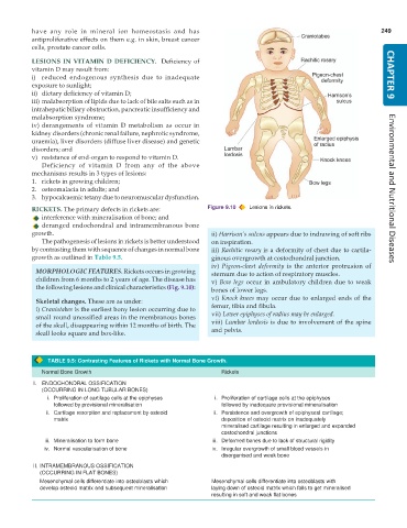
RICKETS. The primary defects in rickets are: Figure 9.10 Lesions in rickets.
interference with mineralisation of bone; and
deranged endochondral and intramembranous bone
growth. ii) Harrison’s sulcus appears due to indrawing of soft ribs
The pathogenesis of lesions in rickets is better understood on inspiration.
by contrasting them with sequence of changes in normal bone iii) Rachitic rosary is a deformity of chest due to cartila-
growth as outlined in Table 9.5. ginous overgrowth at costochondral junction.
iv) Pigeon-chest deformity is the anterior protrusion of
MORPHOLOGIC FEATURES. Rickets occurs in growing sternum due to action of respiratory muscles.
children from 6 months to 2 years of age. The disease has v) Bow legs occur in ambulatory children due to weak
the following lesions and clinical characteristics (Fig. 9.10): bones of lower legs.
vi) Knock knees may occur due to enlarged ends of the
Skeletal changes. These are as under:
i) Craniotabes is the earliest bony lesion occurring due to femur, tibia and fibula.
small round unossified areas in the membranous bones vii) Lower epiphyses of radius may be enlarged.
of the skull, disappearing within 12 months of birth. The viii) Lumbar lordosis is due to involvement of the spine
skull looks square and box-like. and pelvis.
TABLE 9.5: Contrasting Features of Rickets with Normal Bone Growth.
Normal Bone Growth Rickets
I. ENDOCHONDRAL OSSIFICATION
(OCCURRING IN LONG TUBULAR BONES)
i. Proliferation of cartilage cells at the epiphyses i. Proliferation of cartilage cells at the epiphyses
followed by provisional mineralisation followed by inadequate provisional mineralisation
ii. Cartilage resorption and replacement by osteoid ii. Persistence and overgrowth of epiphyseal cartilage;
matrix deposition of osteoid matrix on inadequately
mineralised cartilage resulting in enlarged and expanded
costochondral junctions
iii. Mineralisation to form bone iii. Deformed bones due to lack of structural rigidity
iv. Normal vascularisation of bone iv. Irregular overgrowth of small blood vessels in
disorganised and weak bone
II. INTRAMEMBRANOUS OSSIFICATION
(OCCURRING IN FLAT BONES)
Mesenchymal cells differentiate into osteoblasts which Mesenchymal cells differentiate into osteoblasts with
develop osteoid matrix and subsequent mineralisation laying down of osteoid matrix which fails to get mineralised
resulting in soft and weak flat bones

