


Page 264 - Textbook of Pathology, 6th Edition
P. 264
248
            ii) Squamous metaplasia of pancreatic ductal epithelium may
            lead to obstruction and cystic dilatation.
            iii) Squamous metaplasia of urothelium of the pelvis of kid-
            ney may predispose to pyelonephritis and perhaps to
            renal calculi.
            iv) Long-standing metaplasia may cause progression to
            anaplasia under certain circumstances.
            v) Bone growth in vitamin A deficient animals is retarded.
            vi) Immune dysfunction may occur due to damaged barrier
     SECTION I
            epithelium and compromised immune defenses.
            vii) Pregnant women may have increased risk of maternal
            infection, mortality and impaired embryonic develop-
            ment.
           HYPERVITAMINOSIS A. Very large doses of vitamin A
           can produce toxic manifestations in children as well as in
           adults. These may be acute or chronic.
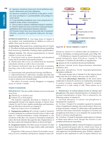
           Acute toxicity. This results from a single large dose of vitamin  Figure 9.9  Normal metabolism of vitamin D.
           A. The effects include neurological manifestations resembling
           brain tumour e.g. headache, vomiting, stupor, papilloedema.
                                                               dihydroxy vitamin D or calcitriol) after its metabolism  in
           Chronic toxicity. The clinical manifestations of chronic  the liver and kidney for being functionally active (Fig. 9.9).
           vitamin A excess are as under:                         1, 25-dihydroxy vitamin D (calcitriol) is 5-10 times more
           i) Neurological such as severe headache and disordered  potent biologically than 25-hydroxy vitamin D. The
           vision due to increased intracranial pressure.      production of calcitriol by the kidney is regulated by:
           ii) Skeletal pains due to loss of cortical bone by increased  plasma levels of calcitriol (hormonal feedback);
           osteoclastic activity as well as due to exostosis.
           iii) Cutaneous involvement may be in the form of pruritus,  plasma calcium levels (hypocalcaemia stimulates
           fissuring, sores at the corners of mouth and coarseness of  synthesis); and
           hair.                                                  plasma phosphorus levels (hypophosphataemia
           iv) Hepatomegaly with parenchymal damage and fibrosis.  stimulates synthesis).
     General Pathology and Basic Techniques
           v) Hypercarotenaemia is yellowness of palms and skin due  The main storage site of vitamin D is the adipose tissue
           to excessive intake of β-carotene containing foods like carrots  rather than the liver which is the case with vitamin A.
           or due to inborn error of metabolism.                  The main  physiologic functions of the most active
              The effects of toxicity usually disappear on stopping  metabolite of vitamin D, calcitriol, are mediated by its binding
           excess of vitamin A intake.                         to nuclear receptor superfamily, vitamin D receptor,
                                                               expressed on a wide variety of cells. These actions are as
           Vitamin D (Calcitriol)                              under:
           PHYSIOLOGY. This fat-soluble vitamin exists in 2 activated  1. Maintenance of normal plasma levels of calcium and
           sterol forms:                                       phosphorus. The major essential function of vitamin D is to
              Vitamin D  or calciferol; and                    promote mineralisation of bone. This is achieved by the
                      2
              Vitamin D  or cholecalciferol.                   following actions of vitamin D:
                      3
              The material originally described as vitamin D  was  i) Intestinal absorption of calcium and phosphorus is
                                                        1
           subsequently found to be impure mixture of sterols. Since  stimulated by vitamin D.
           vitamin D  and D  have similar metabolism and functions,  ii) On bones. Vitamin D is normally required for minerali-
                   2
                          3
           they are therefore referred to as vitamin D.        sation of epiphyseal cartilage and osteoid matrix. However,
              There are 2 main sources of vitamin D:           in hypocalcaemia, vitamin D collaborates with parathyroid
           i) Endogenous synthesis. 80% of body’s need of vitamin D  hormone and causes osteoclastic resorption of calcium and
           is met by endogenous synthesis from the action of ultraviolet  phosphorus from bone so as to maintain the normal blood
           light on 7-dehydrocholesterol widely distributed in oily  levels of calcium and phosphorus.
           secretions of the skin. The vitamin so formed by irradiation  iii) On kidneys. Vitamin D stimulates reabsorption of calcium
           enters the body directly through the skin. Pigmentation of  at distal renal tubular level, though this function is also
           the skin reduces the beneficial effects of ultraviolet light.  parathyroid hormone-dependent.
           ii) Exogenous sources. The other source of vitamin D is diet  2. Antiproliferative effects. Vitamin D receptor is expressed
           such as deep sea fish, fish oil, eggs, butter, milk, some plants  on the parathyroid gland cells by which active form of
           and grains.                                         vitamin D causes antiproliferative action on parathyroid cells
              Irrespective of the source of vitamin D, it must be conver-  and suppresses the parathormone gene. Besides, vitamin D
           ted to its active metabolites (25-hydroxy vitamin D and 1,25-  receptor is also expressed on cells of organs which do not

