


Page 314 - Textbook of Pathology, 6th Edition
P. 314
298 ii) Inadequate intake. Inadequate intake of iron is prevalent  4. INFANTS AND CHILDREN. Iron deficiency anaemia
           in women of lower economic status. Besides diet deficient in  is fairly common during infancy and childhood with a peak
           iron, other factors such as anorexia, impaired absorption and  incidence at 1-2 years of age. The principal cause for anaemia
           diminished bioavailability may act as contributory factors.  at this age is increased demand of iron which is not met by
           iii) Increased requirements. During pregnancy and   the inadequate intake of iron in the diet. Normal full-term
           adolescence, the demand of body for iron is increased. During  infant has sufficient iron stores for the first 4-6 months of
           a normal pregnancy, about 750 mg of iron may be siphoned  life, while premature infants have inadequate reserves
           off from the mother—about 400 mg to the foetus, 150 mg to  because iron stores from the mother are mainly laid down
           the placenta, and 200 mg is lost at parturition and lactation.  during the last trimester of pregnancy. Therefore, unless the
           If several pregnancies occur at short intervals, iron deficiency  infant is given supplemental feeding of iron or iron-
           anaemia certainly follows.                          containing foods, iron deficiency anaemia develops.
           2. POST-MENOPAUSAL FEMALES.   Though the            Clinical Features
           physiological demand for iron decreases after cessation of
           menstruation, iron deficiency anaemia may develop in post-  As already mentioned, iron deficiency anaemia is much more
                                                               common in women between the age of 20 and 45 years than
           menopausal women due to chronic blood loss. Following are  in men; at periods of active growth in infancy, childhood
           among the important causes during these years:      and adolescence; and is also more frequent in premature
           i) Post-menopausal uterine bleeding due to carcinoma of the  infants. Initially, there are usually no clinical abnormalities.
           uterus.                                             But subsequently, in addition to features of the underlying
     SECTION II
           ii) Bleeding from the alimentary tract such as due to carcinoma  disorder causing the anaemia, the clinical consequences of
           of stomach and large bowel and hiatus hernia.       iron deficiency manifest in 2 ways—anaemia itself and
           3. ADULT MALES.  It is uncommon for adult males to  epithelial tissue changes.
           develop iron deficiency anaemia in the presence of normal  1. ANAEMIA.  The onset of iron deficiency anaemia is
           dietary iron content and iron absorption. The vast majority  generally slow. The usual symptoms are weakness, fatigue,
           of cases of iron deficiency anaemia in adult males are due to  dyspnoea on exertion, palpitations and pallor of the skin,
           chronic blood loss. The cause for chronic haemorrhage may  mucous membranes and sclerae. Older patients may develop
           lie at one of the following sites:                  angina and congestive cardiac failure. Patients may have
           i) Gastrointestinal tract is the usual source of bleeding which  unusual dietary cravings such as pica. Menorrhagia is a
           may be due to peptic ulcer, haemorrhoids, hookworm  common symptom in iron deficient women.
           infestation, carcinoma of stomach and large bowel,
           oesophageal varices, hiatus hernia, chronic aspirin ingestion  2. EPITHELIAL  TISSUE  CHANGES.  Long-standing
                                                               chronic iron deficiency anaemia causes epithelial tissue
           and ulcerative colitis. Other causes in the GIT are  changes in some patients. The changes occur in the nails
           malabsorption and following gastrointestinal surgery.  (koilonychia or spoon-shaped nails), tongue (atrophic
           ii) Urinary tract e.g. due to haematuria and haemoglobinuria.  glossitis), mouth (angular stomatitis), and oesophagus
           iii) Nose e.g. in repeated epistaxis.               causing dysphagia from development of thin, membranous
           iv) Lungs e.g. in haemoptysis from various causes.  webs at the postcricoid area (Plummer-Vinson syndrome).


     Haematology and Lymphoreticular Tissues


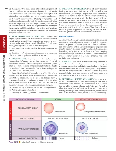
           Figure 12.13  Laboratory findings in iron deficiency anaemia.

