


Page 327 - Textbook of Pathology, 6th Edition
P. 327
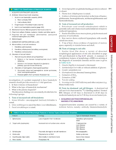
TABLE 12.9: Classification of Haemolytic Anaemias. 4. Serum haptoglobin (α-globulin binding protein) is reduced 311
or absent.
I. ACQUIRED (EXTRACORPUSCULAR) 5. Plasma lactic dehydrogenase is raised.
A. Antibody: Immunohaemolytic anaemias 6. Evidences of intravascular haemolysis in the form of haemo-
1. Autoimmune haemolytic anaemia (AIHA) globinaemia, haemoglobinuria, methaemoglobinaemia and
i) Warm antibody AIHA haemosiderinuria.
ii) Cold antibody AIHA
II. Tests of increased red cell production:
2. Drug-induced immunohaemolytic anaemia
1. Reticulocyte count reveals reticulocytosis which is
3. Isoimmune haemolytic anaemia (page 340)
B. Mechanical trauma: Microangiopathic haemolytic anaemia generally early and is hence most useful initial test of marrow
erythroid hyperplasia.
C. Direct toxic effects: Malaria, bacterial, infection and other agents 2. Routine blood film shows macrocytosis, polychromasia and
D. Acquired red cell membrane abnormalities: paroxysmal presence of normoblasts.
nocturnal haemoglobinuria (PNH)
E. Splenomegaly 3. Bone marrow shows erythroid hyperplasia with usually
raised iron stores.
II. HEREDITARY (INTRACORPUSCULAR) 4. X-ray of bones shows evidence of expansion of marrow
A. Abnormalities of red cell membrane space, especially in tubular bones and skull. CHAPTER 12
1. Hereditary spherocytosis
2. Hereditary elliptocytosis (hereditary ovalocytosis) III. Tests of damage to red cells:
3. Hereditary stomatocytosis 1. Routine blood film shows a variety of abnormal
morphological appearances of red cells described on page
B. Disorders of red cell interior
366 and illustrated in Fig.12.10 already. A summary of
1. Red cell enzyme defects (Enzymopathies) contributory features of morphology of RBCs in arriving at
i) Defects in the hexose monophosphate shunt: G6PD the diagnosis of haemolytic anaemia and its cause is given
deficiency
in Table 12.10.
ii) Defects in the Embden-Meyerhof (or glycolytic)
pathway: pyruvate kinase deficiency 2. Osmotic fragility is increased or decreased.
2. Disorders of haemoglobin (Haemoglobinopathies) 3. Autohaemolysis test with or without addition of glucose.
i) Structurally abnormal haemoglobins: sickle syndromes, 4. Coombs’ antiglobulin test.
other haemoglobinopathies 5. Electrophoresis for abnormal haemoglobins.
ii) Reduced globin chain synthesis: thalassaemias 6. Estimation of HbA .
2
7. Estimation of HbF.
investigations of a patient suspected to have haemolytic 8. Tests for sickling.
anaemia should provide answers to 3 vital questions: 9. Screening test for G6PD deficiency and other enzymes (e.g.
1. Is there evidence of haemolysis? Heinz bodies test).
2. What is the type of haemolytic mechanism? IV.Tests for shortened red cell lifespan. A shortened red
3. What is the precise diagnosis? cell survival is best tested by Cr labelling method. Normal
51
The laboratory findings are conveniently divided into the RBC lifespan of 120 days is shortened to 20-40 days in
following 4 groups: moderate haemolysis and to 5-20 days in severe haemolysis. Introduction to Haematopoietic System and Disorders of Erythroid Series
I. Tests of increased red cell breakdown:
1. Serum bilirubin—unconjugated (indirect) bilirubin is I. ACQUIRED (EXTRACORPUSCULAR)
HAEMOLYTIC ANAEMIAS
raised.
2. Urine urobilinogen is raised but there is no bilirubinuria. Acquired haemolytic anaemias are caused by a variety of
3. Faecal stercobilinogen is raised. extrinsic factors, namely: antibody (immunohaemolytic
TABLE 12.10: Red Cell Morphologic Features in Various Types of Haemolytic Anaemias.
Feature Etiology Type of Haemolytic Anaemia
1. Spherocytes Loss of spectrin from membrane Hereditary spherocytosis
AIHA
2. Target cells (Leptocytes) Increased ratio of surface area: volume Thalassaemias
Liver disease
HbS disease
HbC disease
3. Schistocytes Traumatic damage to red cell membrane Microangiopathy
4. Sickle cells Polymerisation of HbS Sickle syndromes
5. Acanthocytes (Spur cells) Abnormality in membrane lipids Severe liver disease
6. Heinz bodies Precipitated Hb Unstable Hb

