


Page 323 - Textbook of Pathology, 6th Edition
P. 323
307


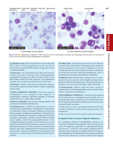
           Figure 12.20  Megaloblastic anaemia. A, PBF showing prominent macrocytosis of red cells and hypersegmented neutrophils. B, Examination  CHAPTER 12
           of bone marrow aspirate showing megaloblastic erythropoiesis.


            iv) Absolute values. The red cell indices reveal an elevated  iii) Other cells. Granulocyte precursors are also affected
            MCV (above 120 fl) proportionate to the severity of  to some extent. Giant forms of metamyelocytes and band
            macrocytosis, elevated MCH (above 50 pg) and normal  cells may be present in the marrow. Megakaryocytes are
            or reduced MCHC.                                     usually present in normal number but may occasionally
            v)Leucocytes. The total white blood cell count may be  be decreased and show abnormal morphology such as
            reduced. Presence of characteristic hypersegmented   hypersegmented nuclei and agranular cytoplasm.
            neutrophils (having more than 5 nuclear lobes) in the  iv)Marrow iron. Prussian blue staining for iron in the
            blood film should raise the suspicion of megaloblastic  marrow shows an increase in the number and size of iron
            anaemia. An occasional myelocyte may also be seen.   granules in the erythroid precursors. Ring sideroblasts are,
            vi)Platelets. Platelet count may be moderately reduced  however, rare. Iron in the reticulum cells is increased.
            in severely anaemic patients. Bizarre forms of platelets  v)Chromosomes.  Marrow cells may show variety of
            may be seen.                                         random chromosomal abnormalities such as chromosome
            2. BONE MARROW FINDINGS. The bone marrow             breaks, centromere spreading etc.                    Introduction to Haematopoietic System and Disorders of Erythroid Series
            examination is very helpful in the diagnosis of      3.BIOCHEMICAL FINDINGS.  In addition to the general
            megaloblastic anaemia. Significant findings of marrow  blood and marrow investigations and specific tests to
            examination are as under (Fig. 12.20,B):
                                                                 determine the cause of deficiency (described below), the
            i) Marrow cellularity. The marrow is hypercellular with  following biochemical abnormalities are observed in cases
            a decreased myeloid-erythroid ratio.                 of megaloblastic anaemia:
            ii) Erythropoiesis.  Erythroid hyperplasia is due to  i) There is rise in serum unconjugated bilirubin and LDH as
            characteristic megaloblastic erythropoiesis. Megaloblasts  a result of ineffective erythropoiesis causing marrow cell
            are abnormal, large, nucleated erythroid precursors,  breakdown.
            having nuclear-cytoplasmic asynchrony i.e. the nuclei are  ii) The serum iron and ferritin may be normal or elevated.
            less mature than the development of cytoplasm. The nuclei
            are large, having fine, reticular and open chromatin that  B. Special Tests for Cause of Specific Deficiency
            stains lightly, while the haemoglobinisation of the
            cytoplasm proceeds normally or at a faster rate i.e. nuclear  In evaluating a patient of megaloblastic anaemia, it is
            maturation lags behind that of cytoplasm (compared from iron  important to determine the specific vitamin deficiency by
            deficiency anaemia in which cytoplasmic maturation lags  assay of vitamin B  and folate. In sophisticated clinical
                                                                                 12
            behind, page 299). Megaloblasts with abnormal mitoses  laboratories, currently automated multiparametric,
            may be seen. Features of ineffective erythropoiesis such  random access analysers are employed based on separa-
            as presence of degenerated erythroid precursors may be  tion techniques by chemiluminescence and enzyme-linked
            present.                                             fluorescence detection systems which have largely

