


Page 331 - Textbook of Pathology, 6th Edition
P. 331
315


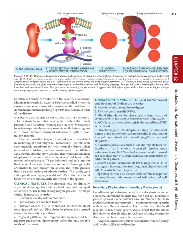
           Figure 12.23  Diagrammatic representation of pathogenesis of hereditary spherocytosis. A, Normal red cell with biconcave surface and normal  CHAPTER 12
           size. B, Red cell membrane as seen in cross section in hereditary spherocytosis. Mutations in membrane proteins—α-spectrin, β-spectrin and
           ankyrin, result in defect in anchoring of  lipid bilayer of the membrane to the underlying cytoskeleton. C, This results in spherical contour and small
           size so as to contain the given volume of haemoglobin in the deformed red cell. D, During passage  through the spleen, these rigid spherical cells
           lose their cell membrane further. This produces a circulating subpopulation of hyperspheroidal spherocytes while splenic macrophages in large
           numbers phagocytose defective red cells causing splenomegaly.


           Spectrin deficiency correlates with the severity of anaemia.  LABORATORY  FINDINGS. The usual haematological
           Mutation in spectrin by recessive inheritance called α−spectrin  and biochemical findings are as under:
           causes more severe form of anaemia, while mutation by  1. Anaemia of mild to moderate degree.
           dominant inheritance forming β-spectrin results in mild form  2. Reticulocytosis, usually 5-20%.
           of the disease.                                       3. Blood film shows the characteristic abnormality of
           2. Ankyrin abnormality.About half the cases of hereditary  erythrocytes in the form of microspherocytes (Fig.12.24).
           spherocytosis have defect in  ankyrin, protein that binds
           protein 3 and spectrin. Homozygous state with recessive  4. MCV is usually normal or slightly decreased but MCHC
                                                                 is increased.
           inheritance pattern has severe anaemia while heterozygotes  5. Osmotic fragility test is helpful in testing the spheroidal
           with more common dominant inheritance pattern have    nature of red cells which lyse more readily in solutions of
           milder anaemia.
              Inherited mutation in spectrin or ankyrin causes defect  low salt concentration i.e.  osmotic fragility is increased
           in anchoring of lipid bilayer cell membrane. Red cells with  (Fig.12.25).
           such unstable membrane but with normal volume, when   6. Autohaemolysis test is similar to osmotic fragility test after
           released in circulation, lose their membrane further, till they  incubation and shows increased spontaneous  Introduction to Haematopoietic System and Disorders of Erythroid Series
           can accommodate the given volume. This results in formation  autohaemolysis (10-15% red cells) as compared to normal
           of spheroidal contour and smaller size of red blood cells,  red cells (less than 4%). Autohaemolysis is correctable by
           termed microspherocytes. These deformed red cells are not  addition of glucose.
           flexible, unlike normal biconcave red cells. These rigid cells  7. Direct Coombs’ (antiglobulin) test  is negative so as to
           are unable to pass through the spleen, and in the process  distinguish this condition from acquired spherocytosis of
           they lose their surface membrane further. This produces a  AIHA in which case it is positive.
           subpopulation of hyperspheroidal red cells in the peripheral  Spherocytes may also be seen in blood film in acquired
           blood which are subsequently destroyed in the spleen.  immune haemolytic anaemia and following red cell
                                                                 transfusion.
           CLINICAL  FEATURES.  The disorder may be clinically
           apparent at any age from infancy to old age and has equal  Hereditary Elliptocytosis (Hereditary Ovalocytosis)
           sex incidence. The family history may be present. The major  Hereditary elliptocytosis or hereditary ovalocytosis is another
           clinical features are as under:                     autosomal dominant disorder involving red cell membrane
           1. Anaemia is usually mild to moderate.             protein spectrin. Some patients have an inherited defect in
           2. Splenomegaly is a constant feature.              erythrocyte membrane protein 4.1 that interconnects spectrin
           3. Jaundice occurs due to increased concentration of  with actin in the cytoskeleton. The disorder is similar in all
           unconjugated (indirect) bilirubin in the plasma (also termed  respects to hereditary spherocytosis except that the blood
           congenital haemolytic jaundice).                    film shows oval or elliptical red cells and is clinically a milder
           4. Pigment gallstones are frequent due to increased bile  disorder than hereditary spherocytosis.
           pigment production. Splenectomy offers the only reliable  Acquired causes of elliptocytosis include iron deficiency
           mode of treatment.                                  and myeloproliferative disorders.

