


Page 328 - Textbook of Pathology, 6th Edition
P. 328
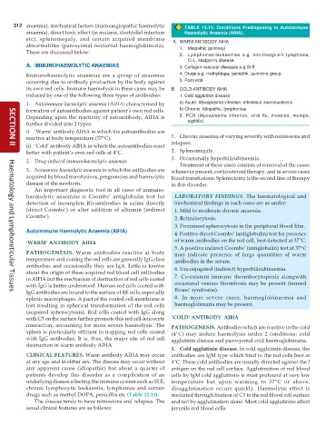
312 anaemia), mechanical factors (microangiopathic haemolytic TABLE 12.11: Conditions Predisposing to Autoimmune
anaemia), direct toxic effect (in malaria, clostridial infection Haemolytic Anaemia (AIHA).
etc), splenomegaly, and certain acquired membrane A. WARM ANTIBODY AIHA
abnormalities (paroxysmal nocturnal haemoglobinuria). 1. Idiopathic (primary)
These are discussed below:
2. Lymphomas-leukaemias e.g. non-Hodgkin’s lymphoma,
CLL, Hodgkin's disease.
A. IMMUNOHAEMOLYTIC ANAEMIAS 3. Collagen vascular diseases e.g SLE
Immunohaemolytic anaemias are a group of anaemias 4. Drugs e.g. methyldopa, penicillin, quinidine group
occurring due to antibody production by the body against 5. Post-viral
its own red cells. Immune haemolysis in these cases may be B. COLD ANTIBODY AIHA
induced by one of the following three types of antibodies: 1. Cold agglutinin disease
1. Autoimmune haemolytic anaemia (AIHA) characterised by a) Acute: Mycoplasma infection, infectious mononucleosis
formation of autoantibodies against patient’s own red cells. b) Chronic: Idiopathic, lymphomas
Depending upon the reactivity of autoantibody, AIHA is 2. PCH (Mycoplasma infection, viral flu, measles, mumps,
further divided into 2 types: syphilis)
i) ‘Warm’ antibody AIHA in which the autoantibodies are
reactive at body temperature (37°C). 1. Chronic anaemia of varying severity with remissions and
ii) ‘Cold’ antibody AIHA in which the autoantibodies react relapses.
SECTION II
better with patient’s own red cells at 4°C. 2. Splenomegaly.
3. Occasionally hyperbilirubinaemia.
2. Drug-induced immunohaemolytic anaemia.
Treatment of these cases consists of removal of the cause
3. Isoimmune haemolytic anaemia in which the antibodies are whenever present, corticosteroid therapy, and in severe cases
acquired by blood transfusions, pregnancies and haemolytic blood transfusions. Splenectomy is the second line of therapy
disease of the newborn. in this disorder.
An important diagnostic tool in all cases of immuno-
haemolytic anaemias is Coombs’ antiglobulin test for LABORATORY FINDINGS. The haematological and
detection of incomplete Rh-antibodies in saline directly biochemical findings in such cases are as under:
(direct Coombs’) or after addition of albumin (indirect 1. Mild to moderate chronic anaemia.
Coombs’). 2. Reticulocytosis.
3. Prominent spherocytosis in the peripheral blood film.
Autoimmune Haemolytic Anaemia (AIHA)
4. Positive direct Coombs’ (antiglobulin) test for presence
‘WARM’ ANTIBODY AIHA of warm antibodies on the red cell, best detected at 37°C.
5. A positive indirect Coombs’ (antiglobulin) test at 37°C
PATHOGENESIS. Warm antibodies reactive at body may indicate presence of large quantities of warm
temperature and coating the red cells are generally IgG class antibodies in the serum.
antibodies and occasionally they are IgA. Little is known 6. Unconjugated (indirect) hyperbilirubinaemia.
about the origin of these acquired red blood cell antibodies
in AIHA but the mechanism of destruction of red cells coated 7. Co-existent immune thrombocytopenia alongwith
with IgG is better understood. Human red cells coated with occasional venous thrombosis may be present (termed
IgG antibodies are bound to the surface of RE cells, especially Evans’ syndrome).
Haematology and Lymphoreticular Tissues
splenic macrophages. A part of the coated cell membrane is 8. In more severe cases, haemoglobinaemia and
lost resulting in spherical transformation of the red cells haemoglobinuria may be present.
(acquired spherocytosis). Red cells coated with IgG along
with C3 on the surface further promote this red cell-leucocyte ‘COLD’ ANTIBODY AIHA
interaction, accounting for more severe haemolysis. The PATHOGENESIS. Antibodies which are reactive in the cold
spleen is particularly efficient in trapping red cells coated (4°C) may induce haemolysis under 2 conditions: cold
with IgG antibodies. It is, thus, the major site of red cell agglutinin disease and paroxysmal cold haemoglobinuria.
destruction in warm antibody AIHA.
1. Cold agglutinin disease. In cold agglutinin disease, the
CLINICAL FEATURES. Warm antibody AIHA may occur antibodies are IgM type which bind to the red cells best at
at any age and in either sex. The disease may occur without 4°C. These cold antibodies are usually directed against the I
any apparent cause (idiopathic) but about a quarter of antigen on the red cell surface. Agglutination of red blood
patients develop this disorder as a complication of an cells by IgM cold agglutinins is most profound at very low
underlying disease affecting the immune system such as SLE, temperature but upon warming to 37°C or above,
chronic lymphocytic leukaemia, lymphomas and certain disagglutination occurs quickly. Haemolytic effect is
drugs such as methyl DOPA, penicillin etc (Table 12.11). mediated through fixation of C3 to the red blood cell surface
The disease tends to have remissions and relapses. The and not by agglutination alone. Most cold agglutinins affect
usual clinical features are as follows: juvenile red blood cells.

