


Page 388 - Textbook of Pathology, 6th Edition
P. 388
372
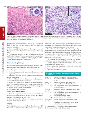
Figure 14.18 Hodgkin’s disease. A, Nodular sclerosis type. There are bands of collagen forming nodules and characteristic lacunar RS cells
(inbox in left figure) . B, Mixed cellularity type. There is admixture of mature lymphocytes, plasma cells, neutrophils and eosinophils and classic RS
SECTION II
cells in the centre of the field (inbox in right figure).
lymph nodes are involved most frequently. Other lymph studied in order to select proper treatment and assess the
node groups like axillary, inguinal and abdominal are prognosis. Ann Arbor staging classification takes into account
involved sometimes. both clinical and pathologic stage of the disease.
2. Approximately half the patients develop splenomegaly The suffix A or B are added to the above stages depen-
during the course of the disease. Liver enlargement too may ding upon whether the three constitutional symptoms (fever,
occur. night sweats and unexplained weight loss exceeding 10% of
3. Constitutional symptoms (type B symptoms) are present normal) are absent (A) or present (B). The suffix E or S are
in 25-40% of patients. The most common is low-grade fever used for extranodal involvement and splenomegaly
with night sweats and weight loss. Other symptoms include respectively (Table 14.10).
fatigue, malaise, weakness and pruritus. For complete staging, a number of other essential diag-
nostic studies are recommended. These are as under:
Other Laboratory Findings 1. Detailed physical examination including sites of nodal
involvement and splenomegaly.
Besides clinical and pathologic findings, there are some 2. Chest radiograph to exclude mediastinal, pleural and
haematologic and immunologic abnormalities in HD. lung parenchymal involvement.
Haematologic abnormalities: 3. CT scan of abdomen and pelvis.
1. A moderate, normocytic and normochromic anaemia is
often present. TABLE 14.10: Ann Arbor Staging Classification of Hodgkin’s
2. Serum iron and TIBC are low but marrow iron stores are Disease.
Haematology and Lymphoreticular Tissues
normal or increased. Stage I I Involvement of a single lymph node region.
3. Marrow infiltration by the disease may produce marrow (A or B) I E Involvement of a single extra-lymphatic organ
failure with leucoerythroblastic reaction. or site.
4. Routine blood counts reveal moderate leukaemoid reaction. Stage II II Involvement of two or more lymph node
Cases with pruritus frequently show peripheral eosinophilia. (A or B) regions on the same side of the diaphragm.
Advanced disease is associated with absolute lymphopenia. II E (or) with localised contiguous involvement of an
5 Platelet count is normal or increased. extranodal organ or site.
6. ESR is invariably elevated. Stage III III Involvement of lymph node regions on both sides
Immunologic abnormalities: (A or B) of the diaphragm.
III E (or) with localised contiguous involvement of an
1. There is progressive fall in immunocompetent T-cells extranodal organ or site.
with defective cellular immunity. There is reversal of CD4: CD8 III S (or) with involvement of spleen.
ratio and anergy to routine skin tests. Ill ES (or) both features of III E and III S .
2. Humoral antibody production is normal in untreated Stage IV IV Multiple or disseminated involvement of one
patients until late in the disease. (A or B) or
more extra-lymphatic organs or tissues with or
Staging without lymphatic involvement.
Following biopsy and histopathologic classification of HD, (A = Asymptomatic; B = Presence of constitutional symptoms;
the extent of involvement of the disease (i.e. staging) is E = Extranodal involvement; S = Splenomegaly).

