


Page 389 - Textbook of Pathology, 6th Edition
P. 389
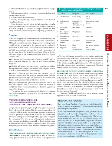
4. Documentation of constitutional symptoms (B symp- TABLE 14.11: Contrasting Features of Hodgkin’s Disease 373
toms). and Non-Hodgkin’s Lymphoma.
5. Laboratory evaluation of complete blood counts, liver and Feature Hodgkin’s Non-Hodgkin’s
kidney function tests.
6. Bilateral bone marrow biopsy. 1. Cell derivation B-cell mostly 90% B
7. Finally, histopathologic documentation of the type of 10% T
Hodgkin’s disease. 2. Nodal involve- Localised, may Disseminated nodal
ment spread to spread
More invasive investigations include lymphangiography contiguous nodes
of lower extremities and staging laparotomy. Staging laparotomy
includes biopsy of selected lymph nodes in the 3. Extranodal Uncommon Common
spread
retroperitoneum, splenectomy and wedge biopsy of the liver.
4. Bone marrow Uncommon Common
involvement
Prognosis
5. Constitutional Common Uncommon
With use of aggressive radiotherapy and chemotherapy, the symptoms
outlook for Hodgkin’s disease has improved significantly. 6. Chromosomal Aneuploidy Translocations,
Although several factors affect the prognosis, two important defects deletions CHAPTER 14
considerations in evaluating its outcome are the extent of 7. Spill-over Never May spread to blood
involvement by the disease (i.e. staging) and the histologic subtype. 8. Prognosis Better Bad
With appropriate treatment, the overall 5 years survival (75-85% cure) (30-40% cure)
rate for stage I and II A is as high as about 100%, while the
advanced stage of the disease may have upto 50% 5-year
survival rate. children or adults and it rapidly transforms into leukaemia.
In cases having leukaemic presentation, extranodal site
Patients with lymphocyte-predominance type of HD tend to
have localised form of the disease and have excellent involvement is early such as lymphadenopathy accompanied
prognosis. with hepatomegaly, splenomegaly, CNS infiltration,
testicular enlargement, and at times cutaneous infiltration.
Nodular sclerosis variety too has very good prognosis but Infections due to cytopenia are present.
those patients with larger mediastinal mass respond poorly
to both chemotherapy and radiotherapy. PRECURSOR T-CELL LYMPHOBLASTIC LEUKAEMIA/
Mixed cellularity type occupies intermediate clinical LYMPHOMA. As the name implies, these cases may present
position between the lymphocyte predominance and the as ALL or as lymphoma. Since the precursor T-cells
lymphocyte-depletion type, but patients with disseminated differentiate in the thymus, this tumour often presents as
disease and systemic manifestations do poorly. mediastinal mass and pleural effusion and progresses rapidly Disorders of Leucocytes and Lymphoreticular Tissues
Lymphocyte-depletion type is usually disseminated at the to develop leukaemia in the blood and bone marrow.
time of diagnosis and is associated with constitutional Clinically, features of bone marrow failure are present which
symptoms. These patients usually have the most aggressive include anaemia, neutropenia and thrombocytopenia.
form of the disease. Lymphadenopathy, hepatosplenomegaly and CNS
The salient features to distinguish Hodgkin’s disease involvement are frequent.
and non-Hodgkin’s lymphoma are summarised in Precursor T-cell lymphoma-leukaemia is, however, more
Table 14.11. aggressive than its B-cell counterpart.
PRECURSOR (IMMATURE) B- AND MORPHOLOGIC FEATURES
T-CELL LEUKAEMIA/LYMPHOMA
(SYNONYM: ACUTE LYMPHOBLASTIC LEUKAEMIA) Precursor B and T-cell ALL/lymphoma are indistinguish-
able on routine morphology. The diagnosis is made by
Lymphoid malignancy originating from precursor series of following investigations:
B or T cell (i.e. pre-B and pre-T) is the most common form of
cancer of children under 4 years of age, together constituting 1. Blood examination. Peripheral blood generally shows
4% of all lymphoid malignancies. Pre-B cell ALL constitutes anaemia and thrombocytopenia, and may show
90% cases while pre-T cell lymphoid malignancies comprise leucopenia-to-normal TLC-to-leucocytosis. DLC shows
the remaining 10%. This group of lymphoid malignancies large number of circulating lymphoblasts having round
arise from more primitive stages of B or T cells but the stage to convoluted nuclei, high nucleo-cytoplasmic ratio and
of differentiation is not related to aggressiveness. Because of absence of cytoplasmic granularity. It is important to
morphologic similarities, both these are presented together. distinguish AML from ALL; the morphologic features of
myeloblasts and lymphoblasts are contrasted in Table 14.1
Clinical Features for comparison (Fig. 14.19). Typical characteristics of
different forms of ALL (L1 to L3) are given in Table 14.6.
PRECURSOR B-CELL LYMPHOBLASTIC LEUKAEMIA/ It is usual to find some ‘smear cells’ in the peripheral blood
LYMPHOMA. Most often, it presents as ALL in children; which represent degenerated leucocytes.
rarely presentation may be in the form of lymphoma in

