


Page 395 - Textbook of Pathology, 6th Edition
P. 395
379


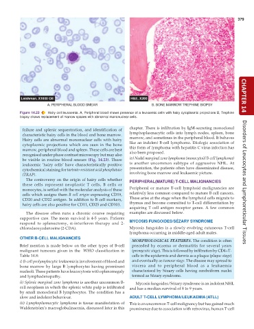
           Figure 14.23  Hairy cell leukaemia. A, Peripheral blood shows presence of a leukaemic cells with hairy cytoplasmic projections B, Trephine  CHAPTER 14
           biopsy shows replacement of marrow spaces with abnormal mononuclear cells.

                                                               chapter. There is infiltration by IgM-secreting monoclonal
            failure and splenic sequestration, and identification of
            characteristic hairy cells in the blood and bone marrow.  lymphoplasmacytic cells into lymph nodes, spleen, bone
            Hairy cells are abnormal mononuclear cells with hairy  marrow, and sometimes in the peripheral blood. It behaves
            cytoplasmic projections which are seen in the bone  like an indolent B-cell lymphoma. Etiologic association of
                                                               this form of lymphoma with hepatitis C virus infection has
            marrow, peripheral blood and spleen. These cells are best  also been proposed.
            recognised under phase contrast microscopy but may also
            be visible in routine blood smears  (Fig. 14.23). These  iv) Nodal marginal zone lymphoma (monocytoid B-cell lymphoma)
            leukaemic ‘hairy cells’ have characteristically positive  is another uncommon subtype of aggressive NHL. At
            cytochemical staining for tartrate-resistant acid phosphatase  presentation, the patients often have disseminated disease,
                                                               involving bone marrow and leukaemic picture.
            (TRAP).
              The controversy on the origin of hairy cells whether  PERIPHERAL(MATURE) T-CELL MALIGNANCIES
            these cells represent neoplastic T cells, B cells or                                                      Disorders of Leucocytes and Lymphoreticular Tissues
            monocytes, is settled with the molecular analysis of these  Peripheral or mature T-cell lymphoid malignancies are
            cells which assigns them B cell origin expressing CD19,  relatively less common compared to mature B cell cancers.
            CD20 and CD22 antigen. In addition to B cell markers,  These arise at the stage when the lymphoid cells migrate to
            hairy cells are also positive for CD11, CD25 and CD103.  thymus and become committed to T-cell differentiation by
                                                               acquiring T cell antigen receptor genes. A few common
              The disease often runs a chronic course requiring  examples are discussed below.
           supportive care. The mean survival is 4-5 years. Patients
           respond to splenectomy,  α-interferon therapy and 2-  MYCOSIS FUNGOIDES/SÉZARY SYNDROME
           chlorodeoxyadenosine (2-CDA).                       Mycosis fungoides is a slowly evolving cutaneous T-cell
                                                               lymphoma occurring in middle-aged adult males.
           OTHER B-CELL MALIGNANCIES
                                                                 MORPHOLOGICAL FEATURES. The condition is often
           Brief mention is made below on the other types of B-cell  preceded by eczema or dermatitis for several years
           malignant tumours given in the  WHO classification in  (premycotic stage). This is followed by infiltration by CD4+T-
           Table 14.8:                                           cells in the epidermis and dermis as a plaque (plaque stage)
           i) B-cell prolymphocytic leukaemia is involvement of blood and  and eventually as tumour stage. The disease may spread to
           bone marrow by large B lymphocytes having prominent   viscera and to peripheral blood as a leukaemia
           nucleoli. These patients have leucocytosis with splenomegaly  characterised by Sézary cells having cerebriform nuclei
           and lymphadenopathy.                                  termed as Sézary syndrome.
           ii) Splenic marginal zone lymphoma is another uncommon B-  Mycosis fungoides/Sézary syndrome is an indolent NHL
           cell neoplasm in which the splenic white pulp is infiltrated  and has a median survival of 8 to 9 years.
           by small monoclonal B lymphocytes. The condition has a
           slow and indolent behaviour.                        ADULT T-CELL LYMPHOMA/LEUKAEMIA (ATLL)
           iii) Lymphoplasmacytic lymphoma is tissue manifestation of  This is an uncommon T-cell malignancy but has gained much
           Waldenström’s macroglobulinaemia, discussed later in this  prominence due to association with retrovirus, human T-cell

