


Page 392 - Textbook of Pathology, 6th Edition
P. 392
376


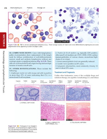
           Figure 14.20  PBF in chronic lymphocytic leukaemia (CLL). There is large excess of mature and small differentiated lymphocytes and some
           degenerated forms appearing as bare smudged nuclei.
     SECTION II


            III. LYMPH NODE BIOPSY. Cases with lymphadeno-       2.  Positive for B-cell markers e.g. typically CD5 positive;
            pathy at presentation show replacement of the lymph  other pan-B cell markers are CD19, CD20, CD23, surface
            node by diffuse proliferation of well-differentiated,  immunoglobulins of various classes, monoclonal light
            mature, small and uniform lymphocytes without any    chains (λ or κ type).
            cytologic atypia or significant mitoses (Fig. 14.21,B). These  3. Serum immunoglobulin levels are generally reduced.
            cells are of monoclonal B-cell origin having immunologic  4. Coombs’ test is positive in 20% cases.
            features of mantle zone B-cells.                     5. Cytogenetic abnormalities, most commonly trisomy 12
            IV. OTHER INVESTIGATIONS.  These include the         seen in about 25% cases.
            following:
            1. Erythrocyte rosette test with mouse red cells is positive  Treatment and Prognosis
            in more than 95% of cases indicating that CLL is a  Unlike other leukaemias, none of the available drugs and
            monoclonal B cell neoplasm.                        radiation therapy are capable of eradicating CLL and induce


     Haematology and Lymphoreticular Tissues


           Figure 14.21  Prototypes of non-Hodgkin’s
           lymphoma—small lymphocytic lymphoma SLL/
           CLL (B) and follicular lymphoma (C) contrasted
           with structure of normal lymph node (A).

