


Page 456 - Textbook of Pathology, 6th Edition
P. 456
440                                                        1. RHEUMATIC ENDOCARDITIS. Endocardial lesions
                                                                 of RF may involve the valvular and mural endocardium,
                                                                 causing  rheumatic valvulitis and  mural endocarditis,
                                                                 respectively. Rheumatic valvulitis is chiefly responsible
                                                                 for the major cardiac manifestations in chronic RHD.
                                                                 RHEUMATIC VALVULITIS. Grossly, the valves in acute
                                                                 RF show thickening and loss of translucency of the valve
                                                                 leaflets or cusps. This is followed by the formation of
                                                                 characteristic, small (1 to 3 mm in diameter), multiple,
                                                                 warty  vegetations or  verrucae, chiefly along the line of
                                                                 closure of the leaflets and cusps. These tiny vegetations
                                                                 are almost continuous so that the free margin of the cusps
                                                                 or leaflets appears as a rough and irregular ridge. The
                                                                 vegetations in RF appear grey-brown, translucent and are
                                                                 firmly attached so that they are not likely to get detached
                                                                 to form emboli, unlike the friable vegetations of infective
                                                                 endocarditis (page 445).
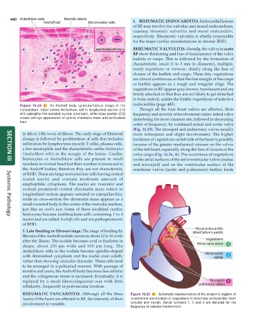
           Figure 16.24  An Aschoff body (granulomatous stage) in the
           myocardium. Inbox shows Anitschkow cell in longitudinal section (LS)  Though all the four heart valves are affected, their
           with caterpillar-like serrated nuclear chromatin, while cross section (CS)  frequency and severity of involvement varies: mitral valve
           shows owl-eye appearance of central chromatin mass and perinuclear  alone being the most common site, followed in decreasing
           halo.
                                                                 order of frequency, by combined mitral and aortic valve
                                                                 (Fig. 16.25). The tricuspid and pulmonary valves usually
            in 4th to 13th week of illness. The early stage of fibrinoid  show infrequent and slight involvement. The higher
            change is followed by proliferation of cells that includes  incidence of vegetations on left side of the heart is possibly
            infiltration by lymphocytes (mostly T cells), plasma cells,  because of the greater mechanical stresses on the valves
            a few neutrophils and the characteristic cardiac histiocytes  of the left heart, especially along the line of closure of the
            (Anitschkow cells) at the margin of the lesion. Cardiac  valve cusps (Fig. 16.26, A). The occurrence of vegetations
            histiocytes or Anitschkow cells are present in small  on the atrial surfaces of the atrioventricular valves (mitral
     SECTION III
            numbers in normal heart but their number is increased in  and tricuspid) and on the ventricular surface of the
            the Aschoff bodies; therefore they are not characteristic  semilunar valves (aortic and pulmonary) further lends
            of RHD. These are large mononuclear cells having central
            round nuclei and contain moderate amount of
            amphophilic cytoplasm. The nuclei are vesicular and
            contain prominent central chromatin mass which in
            longitudinal section appears serrated or caterpillar-like,
            while in cross-section the chromatin mass appears as a
            small rounded body in the centre of the vesicular nucleus,
            just like an owl’s eye. Some of these modified cardiac
            histiocytes become multinucleate cells containing 1 to 4
     Systemic Pathology
            nuclei and are called Aschoff cells and are pathognomonic
            of RHD.
            3. Late (healing or fibrous) stage. The stage of healing by
            fibrosis of the Aschoff nodule occurs in about 12 to 16 weeks
            after the illness. The nodule becomes oval or fusiform in
            shape, about 200  μm wide and 600  μm long. The
            Anitschkow cells in the nodule become spindle-shaped
            with diminished cytoplasm and the nuclei stain solidly
            rather than showing vesicular character. These cells tend
            to be arranged in a palisaded manner. With passage of
            months and years, the Aschoff body becomes less cellular
            and the collagenous tissue is increased. Eventually, it is
            replaced by a small fibrocollagenous scar with little
            cellularity, frequently in perivascular location.
            RHEUMATIC PANCARDITIS. Although all the three      Figure 16.25  Schematic representation of the anatomic regions of
            layers of the heart are affected in RF, the intensity of their  involvement and location of vegetations in rheumatic endocarditis (both
            involvement is variable.                           valvular and mural). Serial numbers 1, 2 and 3 are denoted for the
                                                               frequency of valvular involvement.

