


Page 451 - Textbook of Pathology, 6th Edition
P. 451
435


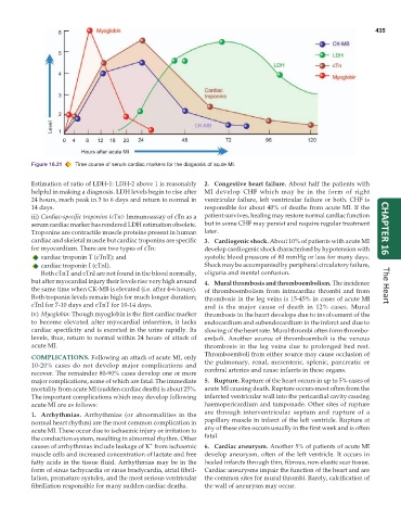
           Figure 16.21  Time course of serum cardiac markers for the diagnosis of acute MI.

           Estimation of ratio of LDH-1: LDH-2 above 1 is reasonably  2. Congestive heart failure. About half the patients with
           helpful in making a diagnosis. LDH levels begin to rise after  MI develop CHF which may be in the form of right
           24 hours, reach peak in 3 to 6 days and return to normal in  ventricular failure, left ventricular failure or both. CHF is
           14 days.                                            responsible for about 40% of deaths from acute MI. If the
           iii) Cardiac-specific troponins (cTn): Immunoassay of cTn as a  patient survives, healing may restore normal cardiac function
           serum cardiac marker has rendered LDH estimation obsolete.  but in some CHF may persist and require regular treatment
           Troponins are contractile muscle proteins present in human  later.                                         CHAPTER 16
           cardiac and skeletal muscle but cardiac troponins are specific  3. Cardiogenic shock. About 10% of patients with acute MI
           for myocardium. There are two types of cTn:         develop cardiogenic shock characterised by hypotension with
              cardiac troponin T (cTnT); and                   systolic blood pressure of 80 mmHg or less for many days.
              cardiac troponin I (cTnI).                       Shock may be accompanied by peripheral circulatory failure,
              Both cTnT and cTnI are not found in the blood normally,  oliguria and mental confusion.
           but after myocardial injury their levels rise very high around  4. Mural thrombosis and thromboembolism. The incidence
           the same time when CK-MB is elevated (i.e. after 4-6 hours).  of thromboembolism from intracardiac thrombi and from  The Heart
           Both troponin levels remain high for much longer duration;  thrombosis in the leg veins is 15-45% in cases of acute MI
           cTnI for 7-10 days and cTnT for 10-14 days.         and is the major cause of death in 12% cases. Mural
           iv) Myoglobin: Though myoglobin is the first cardiac marker  thrombosis in the heart develops due to involvement of the
           to become elevated after myocardial infarction, it lacks  endocardium and subendocardium in the infarct and due to
           cardiac specificity and is excreted in the urine rapidly. Its  slowing of the heart rate. Mural thrombi often form thrombo-
           levels, thus, return to normal within 24 hours of attack of  emboli. Another source of thromboemboli is the venous
           acute MI.                                           thrombosis in the leg veins due to prolonged bed rest.
                                                               Thromboemboli from either source may cause occlusion of
           COMPLICATIONS. Following an attack of acute MI, only
           10-20% cases do not develop major complications and  the pulmonary, renal, mesenteric, splenic, pancreatic or
           recover. The remainder 80-90% cases develop one or more  cerebral arteries and cause infarcts in these organs.
           major complications, some of which are fatal. The immediate  5. Rupture. Rupture of the heart occurs in up to 5% cases of
           mortality from acute MI (sudden cardiac death) is about 25%.  acute MI causing death. Rupture occurs most often from the
           The important complications which may develop following  infarcted ventricular wall into the pericardial cavity causing
           acute MI are as follows:                            haemopericardium and tamponade. Other sites of rupture
                                                               are through interventricular septum and rupture of a
           1. Arrhythmias. Arrhythmias (or abnormalities in the
           normal heart rhythm) are the most common complication in  papillary muscle in infarct of the left ventricle. Rupture at
           acute MI. These occur due to ischaemic injury or irritation to  any of these sites occurs usually in the first week and is often
           the conduction system, resulting in abnormal rhythm. Other  fatal.
                                               +
           causes of arrhythmias include leakage of K  from ischaemic  6. Cardiac aneurysm. Another 5% of patients of acute MI
           muscle cells and increased concentration of lactate and free  develop aneurysm, often of the left ventricle. It occurs in
           fatty acids in the tissue fluid. Arrhythmias may be in the  healed infarcts through thin, fibrous, non-elastic scar tissue.
           form of sinus tachycardia or sinus bradycardia, atrial fibril-  Cardiac aneurysms impair the function of the heart and are
           lation, premature systoles, and the most serious ventricular  the common sites for mural thrombi. Rarely, calcification of
           fibrillation responsible for many sudden cardiac deaths.  the wall of aneurysm may occur.

