


Page 457 - Textbook of Pathology, 6th Edition
P. 457
441


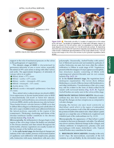
                                                         Figure 16.26  Rheumatic valvulitis. A, Location of vegetations on the valves
                                                         of the left heart. The location of vegetations on mitral valve (left upper diagram) is
                                                         shown as viewed from the left atrium, while the vegetations on aortic valve (left
                                                         lower diagram) are shown as seen from the left ventricular surface.  B, Opened up
                                                         chambers and valves of the left heart show irregularly scarred mitral valve leaving
                                                         a fish-mouth or buttonhole opening between its two cusps (black arrow). The free
                                                         surface and margin of the mitral valve shows tiny firm granular vegetations (white
                                                         arrow).


            support to the role of mechanical pressure on the valves  polymorphs. Occasionally, Aschoff bodies with central
            in the pathogenesis of vegetations.                  foci of fibrinoid necrosis and surrounded by palisade of
               The chronic stage of RHD is characterised by      cardiac histiocytes are seen, but more often the cellular  CHAPTER 16
            permanent deformity of one or more valves, especially  infiltration is diffuse in acute stage of RF. Vegetations
            the mitral (in 98% cases alone or along with other valves)  present at the free margins of cusps appear as eosinophilic,
            and aortic. The approximate frequency of deformity of  tiny structures mainly consisting of fibrin with
            various valves is as under:                          superimposed platelet-thrombi and do not contain
               Mitral alone = 37% cases.                         bacteria (Fig. 16.27, A).
               Mitral + aortic = 27% cases.                      ii) In the healed (chronic) stage, the vegetations have
               Mitral + aortic + tricuspid = 22% cases.          undergone organisation. The valves show diffuse      The Heart
               Mitral + tricuspid = 11% cases.                   thickening as a result of fibrous tissue with hyalinisation,
               Aortic alone = 2%.                                and often calcification. Vascularisation of the valve cusps
               Mitral + aortic + tricuspid + pulmonary = less than  may still be evident in the form of thick-walled blood
            1% cases.                                            vessels with narrowed lumina  (Fig. 16.27, B). Typical
               Thus, mitral valve is almost always involved in RHD.  Aschoff bodies are rarely seen in the valves at this stage.
            Gross appearance of chronic healed mitral valve in RHD  RHEUMATIC MURAL ENDOCARDITIS. Mural endo-
            is characteristically ‘fish mouth’ or ‘button hole’ stenosis.  cardium may also show features of rheumatic carditis
            Mitral stenosis and insufficiency are commonly combined  though the changes are less conspicuous as compared to
            in chronic RHD; calcific aortic stenosis may also be found.  valvular changes.
            These healed chronic valvular lesions in RHD occur due  Grossly, the lesions are seen most commonly as
            to diffuse fibrocollagenous thickening and calcification of  MacCallum’s patch which is the region of endocardial
            the valve cusps or leaflets which cause adhesions between  surface in the posterior wall of the left atrium just above
            the lateral portions, especially in the region of the  the posterior leaflet of the mitral valve. MacCallum’s patch
            commissures. Thickening, shortening and fusion of the  appears as a map-like area of thickened, roughened and
            chordae tendineae further contribute to the chronic  wrinkled part of the endocardium (see Fig. 16.25).
            valvular lesions (Fig. 16.26, B).                    Microscopically, the appearance of MacCallum’s patch
            Microscopically, the inflammatory changes begin in the  is similar to that seen in rheumatic valvulitis. The affected
            region of the valve rings (where the leaflets are attached  area shows oedema, fibrinoid change in the collagen, and
            to the fibrous annulus) and then extend throughout the  cellular infiltrate of lymphocytes, plasma cells and
            entire leaflet, whereas vegetations are usually located on  macrophages with many Anitschkow cells. Typical
            the free margin of the leaflets and cusps.           Aschoff bodies may sometimes be found.
            i) In the early (acute) stage, the histological changes are
            oedema of the valve leaflet, presence of increased number  2. RHEUMATIC MYOCARDITIS. Grossly, in the early
            of capillaries and infiltration with lymphocytes, plasma  (acute)  stage, the myocardium, especially of the left
            cells, histiocytes with many Anitschkow cells and a few  ventricle, is soft and flabby. In the intermediate stage, the

