


Page 458 - Textbook of Pathology, 6th Edition
P. 458
442


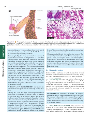
           Figure 16.27  Rheumatic heart disease. A, Microscopic structure of the rheumatic valvulitis and a vegetation on the cusp of mitral valve in
           sagittal section. B, Section of the myocardium shows healed Aschoff nodules in the interstitium having collagen, sparse cellularity, a multinucleate
           giant cell and Anitschkow cells. Inbox shows an Anitschkow cell in cross section (CS) and in longitudinal section (LS).


            interstitial tissue of the myocardium shows small foci of  layers of the pericardium form fibrous adhesions resulting
            necrosis. Later, tiny pale foci of the Aschoff bodies may  in chronic adhesive pericarditis.
            be visible throughout the myocardium.                Microscopically, fibrin is identified on the surfaces. The
            Microscopically, the most characteristic feature of  subserosal connective tissue is infiltrated by lymphocytes,
            rheumatic myocarditis is the presence of distinctive  plasma cells, histiocytes and a few neutrophils.
     SECTION III
            Aschoff bodies. These diagnostic nodules are scattered  Characteristic Aschoff bodies may be seen which later
            throughout the interstitial tissue of the myocardium and  undergo organisation and fibrosis. Organisation of the
            are most frequent in the interventricular septum, left  exudate causes fibrous adhesions between the visceral and
            ventricle and left atrium. Derangements of the conduction  parietal surfaces of the pericardial sac and obliterates the
            system may, thus, be present. The Aschoff bodies are best  pericardial cavity.
            identified in the intermediate stage when they appear as
            granulomas with central fibrinoid necrosis and are  B. Extracardiac Lesions
            surrounded by palisade of Anitschkow cells and     Patients of the syndrome of acute rheumatism develop
            multinucleate Aschoff cells. There is infiltration by  lesions in connective tissue elsewhere in the body, chiefly
            lymphocytes, plasma cells and some neutrophils. In the  the joints, subcutaneous tissue, arteries, brain and lungs.
            late stage, the Aschoff bodies are gradually replaced by
     Systemic Pathology
            small fibrous scars in the vicinity of blood vessels and the  1. POLYARTHRITIS. Acute and painful inflammation of
            inflammatory infiltrate subsides. Presence of active  the synovial membranes of some of the joints, especially the
            Aschoff bodies along with old healed lesions is indicative  larger joints of the limbs, is seen in about 90% cases of RF in
            of rheumatic activity.                             adults and less often in children. As pain and swelling
            3. RHEUMATIC PERICARDITIS. Inflammatory            subside in one joint, others tend to get involved, producing
            involvement of the pericardium commonly accompanies  the characteristic  ‘migratory polyarthritis’ involving two or
            RHD.                                               more joints at a time.
            Grossly, the usual finding is fibrinous pericarditis in  Histologically, the changes are transitory. The synovial
            which there is loss of normal shiny pericardial surface due  membrane and the periarticular connective tissue show
            to deposition of fibrin on its surface and accumulation of  hyperaemia, oedema, fibrinoid change and neutrophilic
            slight amount of fibrinous exudate in the pericardial sac.  infiltration. Sometimes, focal lesions resembling Aschoff
            If the parietal pericardium is pulled off from the visceral  bodies are observed. A serous effusion into the joint cavity
            pericardium, the two separated surfaces are shaggy due  is commonly present.
            to thick fibrin covering them. This appearance is often
            likened to ‘bread and butter appearance’ i.e. resembling the  2. SUBCUTANEOUS NODULES. The subcutaneous
            buttered surfaces of two slices in a sandwich when they  nodules of RF occur more often in children than in adult.
            are gently pulled apart. If fibrinous pericarditis fails to  These nodules are small (0.5 to 2 cm in diameter), spherical
            resolve and, instead, undergoes organisation, the two  or ovoid and painless. They are attached to deeper structures

