


Page 452 - Textbook of Pathology, 6th Edition
P. 452
436 7. Pericarditis. Sterile pericarditis appearing on about the  ii) An alternate concept of development of myocardial
           second day is common over transmural infarcts. It is  fibrosis is healing of minute areas of focal myocytolysis—
           characterised by fibrinous pericarditis and may be associated  the myocardial fibres in a small area undergo slow
           with pericardial effusion. Often, it is of no functional  degeneration due to myocardial ischaemia. These fibres lose
           significance and resolves spontaneously.            their myofibrils but nuclei remain intact. These foci are
           8. Postmyocardial infarction syndrome. About 3 to 4% of  infiltrated by macrophages and eventually are replaced by
           patients who suffered from acute MI develop postmyocardial  proliferating fibroblasts and collagen.
           infarction syndrome or Dressler’s syndrome subsequently. It  MORPHOLOGIC FEATURES. Grossly, the heart may be
           usually occurs 1 to 6 weeks after the attack of MI. It is  normal in size or hypertrophied. The left ventricular wall
           characterised by pneumonitis. The symptoms are usually  generally shows foci of grey-white fibrosis in brown
           mild and disappear in a few weeks. The exact pathogenesis  myocardium. Healed scars of previous MI may be present.
           of this syndrome is not known. It may be due to autoimmune  Valves of the left heart may be distorted, thickened and
           reaction as evidenced by circulating anti-heart antibodies in  show calcification. Coronary arteries invariably show
           the serum of these patients. But these antibodies are also pre-  moderate to severe atherosclerosis.
           sent in patients with acute MI who do not develop this  Microscopically, the characteristic features are as follows
           syndrome.
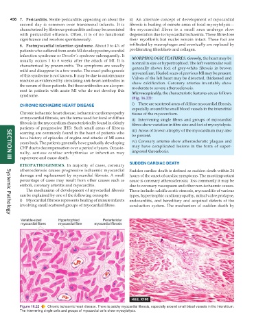
                                                                 (Fig. 16.22):
           CHRONIC ISCHAEMIC HEART DISEASE                       i) There are scattered areas of diffuse myocardial fibrosis,
                                                                 especially around the small blood vessels in the interstitial
           Chronic ischaemic heart disease, ischaemic cardiomyopathy  tissue of the myocardium.
           or myocardial fibrosis, are the terms used for focal or diffuse  ii) Intervening single fibres and groups of myocardial
           fibrosis in the myocardium characteristically found in elderly  fibres show variation in fibre size and foci of myocytolysis.
           patients of progressive IHD. Such small areas of fibrous  iii) Areas of brown atrophy of the myocardium may also
           scarring are commonly found in the heart of patients who  be present.
           have history of episodes of angina and attacks of MI some
           years back. The patients generally have gradually developing  iv) Coronary arteries show atherosclerotic plaques and
           CHF due to decompensation over a period of years. Occasio-  may have complicated lesions in the form of super-
           nally, serious cardiac arrhythmias or infarction may  imposed thrombosis.
           supervene and cause death.
     SECTION III
                                                               SUDDEN CARDIAC DEATH
           ETIOPATHOGENESIS.  In majority of cases, coronary
           atherosclerosis causes progressive ischaemic myocardial  Sudden cardiac death is defined as sudden death within 24
           damage and replacement by myocardial fibrosis. A small  hours of the onset of cardiac symptoms. The most important
           percentage of cases may result from other causes such as  cause is coronary atherosclerosis;  less commonly it may be
           emboli, coronary arteritis and myocarditis.         due to coronary vasospasm and other non-ischaemic causes.
              The mechanism of development of myocardial fibrosis  These include: calcific aortic stenosis, myocarditis of various
           can be explained by one of the following concepts:  types, hypertrophic cardiomyopathy, mitral valve prolapse,
           i) Myocardial fibrosis represents healing of minute infarcts  endocarditis, and hereditary and acquired defects of the
           involving small scattered groups of myocardial fibres.  conduction system. The mechanism of sudden death by

     Systemic Pathology


           Figure 16.22  Chronic ischaemic heart disease. There is patchy myocardial fibrosis, especially around small blood vessels in the interstitium.
           The intervening single cells and groups of myocardial cells show myocytolysis.

