


Page 462 - Textbook of Pathology, 6th Edition
P. 462
446


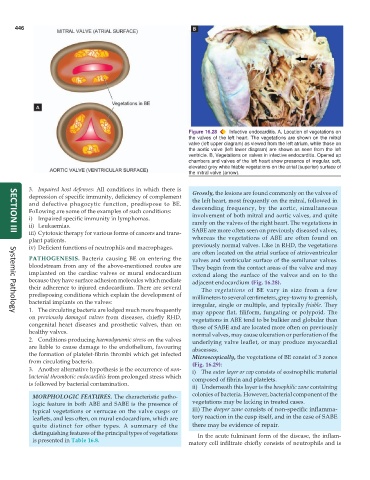
                                                               Figure 16.28  Infective endocarditis. A, Location of vegetations on
                                                               the valves of the left heart. The vegetations are shown on the mitral
                                                               valve (left upper diagram) as viewed from the left atrium, while those on
                                                               the aortic valve (left lower diagram) are shown as seen from the left
                                                               ventricle. B, Vegetations on valves in infective endocarditis. Opened up
                                                               chambers and valves of the left heart show presence of irregular, soft,
                                                               elevated grey white friable vegetations on the atrial (superior) surface of
                                                               the mitral valve (arrow).

           3. Impaired host defenses: All conditions in which there is  Grossly, the lesions are found commonly on the valves of
           depression of specific immunity, deficiency of complement  the left heart, most frequently on the mitral, followed in
           and defective phagocytic function, predispose to BE.  descending frequency, by the aortic, simultaneous
           Following are some of the examples of such conditions:
           i) Impaired specific immunity in lymphomas.           involvement of both mitral and aortic valves, and quite
           ii) Leukaemias.                                       rarely on the valves of the right heart. The vegetations in
           iii) Cytotoxic therapy for various forms of cancers and trans-  SABE are more often seen on previously diseased valves,
     SECTION III
           plant patients.                                       whereas the vegetations of ABE are often found on
           iv) Deficient functions of neutrophils and macrophages.  previously normal valves. Like in RHD, the vegetations
                                                                 are often located on the atrial surface of atrioventricular
           PATHOGENESIS.  Bacteria causing BE on entering the    valves and ventricular surface of the semilunar valves.
           bloodstream from any of the above-mentioned routes are  They begin from the contact areas of the valve and may
           implanted on the cardiac valves or mural endocardium  extend along the surface of the valves and on to the
           because they have surface adhesion molecules which mediate  adjacent endocardium (Fig. 16.28).
           their adherence to injured endocardium. There are several  The vegetations of BE vary in size from a few
           predisposing conditions which explain the development of  millimeters to several centimeters, grey-tawny to greenish,
           bacterial implants on the valves:                     irregular, single or multiple, and typically friable. They
           1. The circulating bacteria are lodged much more frequently  may appear flat, filiform, fungating or polypoid. The
     Systemic Pathology
           on  previously damaged valves from diseases, chiefly RHD,  vegetations in ABE tend to be bulkier and globular than
           congenital heart diseases and prosthetic valves, than on  those of SABE and are located more often on previously
           healthy valves.                                       normal valves, may cause ulceration or perforation of the
           2. Conditions producing haemodynamic stress on the valves  underlying valve leaflet, or may produce myocardial
           are liable to cause damage to the endothelium, favouring  abscesses.
           the formation of platelet-fibrin thrombi which get infected  Microscopically, the vegetations of BE consist of 3 zones
           from circulating bacteria.
                                                                 (Fig. 16.29):
           3. Another alternative hypothesis is the occurrence of non-  i) The outer layer or cap consists of eosinophilic material
           bacterial thrombotic endocarditis from prolonged stress which  composed of fibrin and platelets.
           is followed by bacterial contamination.
                                                                 ii) Underneath this layer is the basophilic zone containing
            MORPHOLOGIC FEATURES. The characteristic patho-      colonies of bacteria. However, bacterial component of the
            logic feature in both ABE and SABE is the presence of  vegetations may be lacking in treated cases.
            typical vegetations or verrucae on the valve cusps or  iii) The deeper zone consists of non-specific inflamma-
            leaflets, and less often, on mural endocardium, which are  tory reaction in the cusp itself, and in the case of SABE
            quite distinct for other types. A summary of the     there may be evidence of repair.
            distinguishing features of the principal types of vegetations  In the acute fulminant form of the disease, the inflam-
            is presented in Table 16.8.                        matory cell infiltrate chiefly consists of neutrophils and is

