


Page 48 - Textbook of Pathology, 6th Edition
P. 48
32 1. Calcium overload.
TABLE 3.1: Common Enzyme Markers of Cell Death.
2. Generation of reactive oxygen radicals (superoxide, H O ,
Enzyme Disease 2 2
hydroxyl radicals).
1. Aspartate aminotransferase Diffuse liver cell necrosis e.g. 3. Subsequent inflammatory reaction.
(AST, SGOT) viral hepatitis, alcoholic liver These are discussed below:
disease
Acute myocardial infarction 1. CALCIUM OVERLOAD. Upon restoration of blood
2. Alanine aminotransferase More specific for diffuse liver supply, the ischaemic cell is further bathed by the blood fluid
(ALT, SGPT) cell damage than AST e.g. that has more calcium ions at a time when the ATP stores of
viral hepatitis the cell are low. This results in further calcium overload on
SECTION I
3. Creatine kinase-MB (CK-MB) Acute myocardial infarction, the already injured cells, triggering lipid peroxidation of the
myocarditis
Skeletal muscle injury membrane causing further membrane damage.
4. Lipase More specific for acute 2. GENERATION OF REACTIVE OXYGEN RADICALS.
pancreatitis Although oxygen is the lifeline of all cells and tissues, its
5. Amylase Acute pancreatitis molecular forms as reactive oxygen radicals or reactive
Sialadenitis
6. Lactic dehydrogenase (LDH) Acute myocardial infarction oxygen species can be most devastating for the cells. In recent
Myocarditis times, free radical-mediated cell injury has been extensively
Skeletal muscle injury studied and a brief account is given below.
7. Cardiac troponin (CTn) Specific for acute myocardial Mechanism of oxygen free radical generation. Normally,
infarction
metabolism of the cell involves generation of ATP by
oxidative process in which biradical oxygen (O ) combines
2
1. From ischaemia to reversible injury. When the period of with hydrogen atom (H) and in the process forms water
ischaemia is of short duration, reperfusion with resupply of (H O). This reaction of O to H O involves ‘four electron
2
2
2
oxygen restores the structural and functional state of the donation’ in four steps involving transfer of one electron at
injured cell i.e. reversible cell injury. each step. Oxygen free radicals are the intermediate chemical
2. From ischaemia to reperfusion injury. When ischaemia is species having an unpaired oxygen in their outer orbit. These
for longer duration, then rather than restoration of structure are generated within mitochondrial inner membrane where
and function of the cell, reperfusion paradoxically cytochrome oxidase catalyses the O to H O reaction. Three
2
2
deteriorates the already injured cell. This is termed intermediate molecules of partially reduced species of oxygen
ischaemia-reperfusion injury. are generated depending upon the number of electrons
General Pathology and Basic Techniques
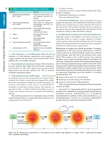
transferred (Fig. 3.8):
3. From ischaemia to irreversible injury. Much longer period Superoxide oxygen (O’ ): one electron
of ischaemia may produce irreversible cell injury during 2
ischaemia itself when so much time has elapsed that neither Hydrogen peroxide (H O ): two electrons
2
2
–
blood flow restoration is helpful nor reperfusion injury can Hydroxyl radical (OH ): three electrons
develop. Cell death in such cases is not attributed to These are generated from enzymatic and non-enzymatic
formation of activated oxygen species. But instead, on reaction as under:
reperfusion there is further marked intracellular excess of 1. Superoxide (O’ ): Superoxide anion O’ may be generated
2
2
sodium and calcium ions due to persistent cell membrane by direct auto-oxidation of O during mitochondrial electron
2
damage. transport reaction. Alternatively, O’ is produced
2
The underlying mechanism of reperfusion injury and free enzymatically by xanthine oxidase and cytochrome P 450 in
radical mediated injury is complex but following three main the mitochondria or cytosol. O’ so formed is catabolised to
2
components are involved in it: produce H O by superoxide dismutase (SOD).
2
2
Figure 3.8 Mechanisms of generation of free radicals by four electron step reduction of oxygen. (SOD = superoxide dismutase;
GSH = glutathione peroxidase).

