


Page 52 - Textbook of Pathology, 6th Edition
P. 52
36


     SECTION I


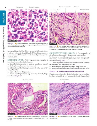
           Figure 3.12  Intracellular hyaline as Russell’s bodies in the plasma
           cells. The cytoplasm shows pink homogeneous globular material due to
           accumulated immunoglobulins.                        Figure 3.13  Extracellular hyaline deposit in leiomyoma uterus. The
                                                               centres of whorls of smooth muscle and connective tissue show pink
                                                               homogeneous hyaline material (connective tissue hyaline).
           are stained by alcian blue. However, epithelial mucin stains
           positively with periodic acid-Schiff (PAS), while connective  CONNECTIVE  TISSUE   MUCIN.  A few examples of
           tissue mucin is PAS negative but is stained positively with  disturbances of connective tissue mucin are as under:
           colloidal iron.                                     1. Mucoid or myxoid degeneration in some tumours e.g.
                                                               myxomas, neurofibromas, fibroadenoma, soft tissue
           EPITHELIAL MUCIN.  Following are some examples of   sarcomas etc (Fig. 3.15) .
           functional excess of epithelial mucin:              2. Dissecting aneurysm of the aorta due to Erdheim’s medial
           1. Catarrhal inflammation of mucous membrane (e.g. of  degeneration and Marfan’s syndrome.
     General Pathology and Basic Techniques
           respiratory tract, alimentary tract, uterus).       3. Myxomatous change in the dermis in myxoedema.
           2. Obstruction of duct leading to mucocele in the oral cavity  4. Myxoid change in the synovium in ganglion on the wrist.
           and gallbladder.
           3. Cystic fibrosis of the pancreas.                 SUBCELLULAR ALTERATIONS IN CELL INJURY
           4. Mucin-secreting tumours (e.g. of ovary, stomach, large  Certain morphologically distinct alterations at subcellular
           bowel etc) (Fig. 3.14) .                            level are noticeable in both acute and chronic forms of cell


           Figure 3.14  Epithelial mucin. Mucinous cystadenoma of the ovary  Figure 3.15  Connective tissue mucin (myxoid change) in
           showing intracytoplasmic mucinous material in the epithelial cells lining  neurofibroma.
           the cyst.

