


Page 49 - Textbook of Pathology, 6th Edition
P. 49
2. Hydrogen peroxide (H O ): H O  is reduced to water  peroxides. This reaction is termed lipid peroxidation. The  33
                                          2
                                       2
                                2
                                   2
           enzymatically by catalase (in the peroxisomes) and  lipid peroxides are decomposed by transition metals such
           glutathione peroxidase GSH (both in the cytosol and  as iron. Lipid peroxidation is propagated to other sites
           mitochondria).                                      causing widespread membrane damage and destruction of
                                    –
           3. Hydroxyl radical (OH ): OH  radical is formed by 2 ways  organelles.
                               –
           in biologic processes—by radiolysis of water and by reaction  ii) Oxidation of proteins. Oxygen-derived free radicals cause  CHAPTER 3
                               ++
           of H O  with ferrous (Fe ) ions; the latter process is termed  cell injury by oxidation of protein macromolecules of the cells,
              2
                 2
           as Fenton reaction.                                 crosslinkages of labile amino acids as well as by fragmen-
                                                               tation of polypeptides directly. The end-result is degradation
           Other oxygen free radicals.  In addition to superoxide, H O 2  of cytosolic neutral proteases and cell destruction.
                                                            2
           and hydroxyl radicals generated during of O to H O
                                                           2
                                                     2
           reaction, a few other more active oxygen free  radicals which  iii) DNA damage. Free radicals cause breaks in the single
           formed in the body are as follows:                  strands of the nuclear and mitochondrial DNA. This results
           i) Release of superoxide free radical in Fenton reaction (see  in cell injury; it may also cause malignant transformation of
           below).                                             cells.
           ii) Nitric oxide (NO), a chemical mediator generated by  iv) Cytoskeletal damage. Reactive oxygen species are also
           various body cells (endothelial cells, neurons, macrophages  known to interact with cytoskeletal elements and interfere
           etc), combines with superoxide and forms peroxynitrate  in mitochondrial aerobic phosphorylation and thus cause
           (ONOO) which is a potent free radical.              ATP depletion.                                         Cell Injury and Cellular Adaptations
           iii)  Halide reagent (chlorine or chloride) released in the  Conditions with free radical injury.  Currently, oxygen-
           leucocytes reacts with superoxide and forms hypochlorous acid  derived free radicals have been known to play an important
           (HOCl) which is a cytotoxic free radical.           role in many forms of cell injury:
           iv) Exogenous sources of free radicals include some environ-  i) Ischaemic reperfusion injury
           mental agents such as tobacco and industrial pollutants.  ii) Ionising radiation by causing radiolysis of water
                                                               iii) Chemical toxicity
           Cytotoxicity of oxygen free radicals.  Free radicals are formed  iv) Chemical carcinogenesis
           in physiologic as well as pathologic processes. Basically,  v) Hyperoxia (toxicity due to oxygen therapy)
           oxygen radicals are unstable and are destroyed spon-  vi) Cellular aging
           taneously. The rate of spontaneous destruction is determined  vii) Killing of microbial agents
           by catalytic action of certain enzymes such as superoxide  viii)  Inflammatory damage
           dismutase (SOD), catalase and glutathione peroxidase. The  ix) Destruction of tumour cells
           net effect of free radical injury in physiologic and disease  x) Atherosclerosis.
           states, therefore, depends upon the rate of free radical
           formation and rate of their elimination.            Antioxidants. Antioxidants are endogenous or exogenous
              However, if not degraded, then free radicals are highly  substances which inactivate the free radicals. These
           destructive to the cell since they have electron-free residue  substances include the following:
           and thus bind to all molecules of the cell; this is termed  Vitamins E, A and C (ascorbic acid)
           oxidative stress.  Out of various free radicals, hydroxyl radical  Sulfhydryl-containing compounds e.g. cysteine and
           is the most reactive species. Free radicals may produce  glutathione.
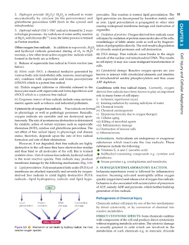
           membrane damage by the following mechanisms (Fig. 3.9):  Serum proteins e.g. ceruloplasmin and transferrin.
           i) Lipid peroxidation. Polyunsaturated fatty acids (PUFA) of  3. SUBSEQUENT INFLAMMATORY EACTION.
           membrane are attacked repeatedly and severely by oxygen-  Ischaemia-reperfusion event is followed by inflammatory
           derived free radicals to yield highly destructive PUFA  reaction. Incoming activated neutrophils utilise oxygen
           radicals—lipid hydroperoxy radicals and lipid hypo-  quickly (oxygen burst) and release a lot of oxygen free radicals.
                                                               Ischaemia is also associated with accumulation of precursors
                                                               of ATP, namely ADP and pyruvate, which further build-up
                                                               generation of free radicals.

                                                               Pathogenesis of Chemical Injury
                                                               Chemicals induce cell injury by one of the two mechanisms:
                                                               by direct cytotoxicity, or by conversion of chemical into
                                                               reactive metabolites.

                                                               DIRECT CYTOTOXIC EFFECTS. Some chemicals combine
                                                               with components of the cell and produce direct cytotoxicity
                                                               without requiring metabolic activation. The cytotoxic damage
           Figure 3.9  Mechanism of cell death by hydroxyl radical, the most  is usually greatest to cells which are involved in the
           reactive oxygen species.                            metabolism of such chemicals e.g. in mercuric chloride

