


Page 51 - Textbook of Pathology, 6th Edition
P. 51
acute and subacute cell injury from various etiologic agents  INTRACELLULAR  HYALINE. Intracellular hyaline is  35
           such as bacterial toxins, chemicals, poisons, burns, high fever,  mainly seen in epithelial cells. A few examples are as follows:
           intravenous administration of hypertonic glucose or saline  1. Hyaline droplets in the proximal tubular epithelial cells in
           etc.                                                cases of excessive reabsorption of plasma proteins.
           PATHOGENESIS. Cloudy swelling results from impaired  2. Hyaline degeneration of rectus abdominalis muscle called
           regulation of sodium and potassium at the level of cell  Zenker’s degeneration, occurring in typhoid fever. The  CHAPTER 3
           membrane. This results in intracellular accumulation of  muscle loses its fibrillar staining and becomes glassy and
           sodium and escape of potassium. This, in turn, leads to rapid  hyaline.
           flow of water into the cell to maintain iso-osmotic conditions  3. Mallory’s hyaline represents aggregates of intermediate
           and hence cellular swelling occurs. In addition, influx of  filaments in the hepatocytes in alcoholic liver cell injury.
           calcium too occurs. Hydropic swelling is an entirely  4. Nuclear or cytoplasmic hyaline inclusions seen in some
           reversible change upon removal of the injurious agent.  viral infections.
                                                               5. Russell’s bodies representing excessive immunoglobulins
            MORPHOLOGIC FEATURES. Grossly, the affected        in the rough endoplasmic reticulum of the plasma cells
            organ such as kidney, liver, pancreas, or heart muscle is  (Fig. 3.12).
            enlarged due to swelling. The cut surface bulges outwards
            and is slightly opaque.                            EXTRACELLULAR HYALINE. Extracellular hyaline is seen
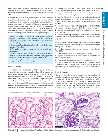
            Microscopically, it is characterised by the following  in connective tissues. A few examples of extracellular hyaline
            features (Fig. 3.11):                              change are as under:                                   Cell Injury and Cellular Adaptations
            i) The cells are swollen and the microvasculature  1. Hyaline degeneration in  leiomyomas of the uterus
            compressed.                                        (Fig. 3.13).
            ii) Small clear vacuoles are seen in the cells and hence  2. Hyalinised old scar of fibrocollagenous tissues.
            the term vacuolar degeneration. These vacuoles represent  3. Hyaline arteriolosclerosis in renal vessels in hypertension
            distended cisternae of the endoplasmic reticulum.  and diabetes mellitus.
            iii) Small cytoplasmic blebs may be seen.          4. Hyalinised glomeruli in chronic glomerulonephritis.
            iv) The nucleus may appear pale.                   5. Corpora amylacea are rounded masses of concentric hya-
                                                               line laminae seen in the prostate in the elderly, in the brain
           Hyaline Change                                      and in the spinal cord in old age, and in old infarcts of the
                                                               lung.
           The word ‘hyaline’ means glassy (hyalos = glass). Hyaline is
           a descriptive histologic term for glassy, homogeneous,  Mucoid Change
           eosinophilic appearance of material in haematoxylin and  Mucus secreted by mucous glands is a combination of
           eosin-stained sections and does not refer to any specific  proteins complexed with mucopolysaccharides. Mucin, a
           substance. Though fibrin and amyloid have hyaline appear-  glycoprotein, is its chief constituent. Mucin is normally
           ance, they have distinctive features and staining reactions  produced by epithelial cells of mucous membranes and
           and can be distinguished from non-specific hyaline material.  mucous glands, as well as by some connective tissues like
           Hyaline change is associated with heterogeneous pathologic  in the umbilical cord. By convention, connective tissue
           conditions. It may be intracellular or extracellular.  mucin is termed myxoid (mucus like). Both types of mucin


           Figure 3.11  Hydropic change kidney. The tubular epithelial cells are distended with cytoplasmic vacuoles while the interstitial vasculature is
           compressed. The nuclei of affected tubules are pale.

