


Page 50 - Textbook of Pathology, 6th Edition
P. 50
34 rane as well as may interact with DNA of the target cell. In
proliferating cells, there is inhibition of DNA replication and
eventual cell death by apoptosis (e.g. epithelial cells). In non-
proliferating cells there is no effect of inhibition of DNA
synthesis and in these cells there is cell membrane damage
followed by cell death by necrosis (e.g. neurons).
MORPHOLOGY OF CELL INJURY
SECTION I
After having discussed the molecular and biochemical
mechanisms of various forms of cell injury, we now turn to
light microscopic morphologic changes of reversible and
irreversible cell injury.
Depending upon the severity of cell injury, degree of
damage and residual effects on cells and tissues are variable.
In general, morphologic changes in various forms of cell
injury can be classified as shown in Table 3.2 and are
discussed below.
MORPHOLOGY OF REVERSIBLE CELL INJURY
In conventional description of morphologic changes, the term
degeneration has been used to denote morphology of
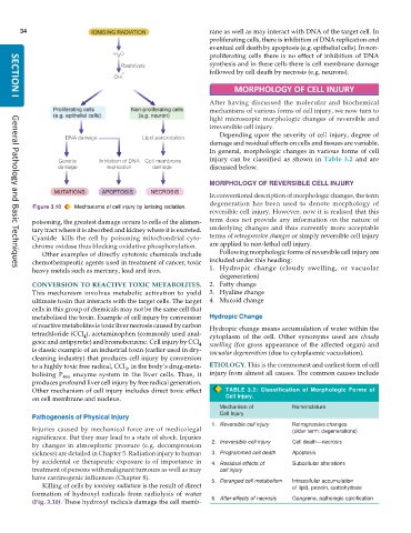
Figure 3.10 Mechanisms of cell injury by ionising radiation.
reversible cell injury. However, now it is realised that this
poisoning, the greatest damage occurs to cells of the alimen- term does not provide any information on the nature of
tary tract where it is absorbed and kidney where it is excreted. underlying changes and thus currently more acceptable
Cyanide kills the cell by poisoning mitochondrial cyto- terms of retrogressive changes or simply reversible cell injury
chrome oxidase thus blocking oxidative phosphorylation. are applied to non-lethal cell injury.
Other examples of directly cytotoxic chemicals include Following morphologic forms of reversible cell injury are
chemotherapeutic agents used in treatment of cancer, toxic included under this heading:
heavy metals such as mercury, lead and iron. 1. Hydropic change (cloudy swelling, or vacuolar
General Pathology and Basic Techniques
degeneration)
CONVERSION TO REACTIVE TOXIC METABOLITES. 2. Fatty change
This mechanism involves metabolic activation to yield 3. Hyaline change
ultimate toxin that interacts with the target cells. The target 4. Mucoid change
cells in this group of chemicals may not be the same cell that
metabolised the toxin. Example of cell injury by conversion Hydropic Change
of reactive metabolites is toxic liver necrosis caused by carbon Hydropic change means accumulation of water within the
tetrachloride (CCl ), acetaminophen (commonly used anal- cytoplasm of the cell. Other synonyms used are cloudy
4
gesic and antipyretic) and bromobenzene. Cell injury by CCl 4 swelling (for gross appearance of the affected organ) and
is classic example of an industrial toxin (earlier used in dry- vacuolar degeneration (due to cytoplasmic vacuolation).
cleaning industry) that produces cell injury by conversion
to a highly toxic free radical, CCl , in the body’s drug-meta- ETIOLOGY. This is the commonest and earliest form of cell
3
bolising P 450 enzyme system in the liver cells. Thus, it injury from almost all causes. The common causes include
produces profound liver cell injury by free radical generation.
Other mechanism of cell injury includes direct toxic effect TABLE 3.2: Classification of Morphologic Forms of
on cell membrane and nucleus. Cell Injury.
Mechanism of Nomenclature
Cell Injury
Pathogenesis of Physical Injury
1. Reversible cell injury Retrogressive changes
Injuries caused by mechanical force are of medicolegal (older term: degenerations)
significance. But they may lead to a state of shock. Injuries
by changes in atmospheric pressure (e.g. decompression 2. Irreversible cell injury Cell death—necrosis
sickness) are detailed in Chapter 5. Radiation injury to human 3. Programmed cell death Apoptosis
by accidental or therapeutic exposure is of importance in 4. Residual effects of Subcellular alterations
treatment of persons with malignant tumours as well as may cell injury
have carcinogenic influences (Chapter 8). 5. Deranged cell metabolism Intracellular accumulation
Killing of cells by ionising radiation is the result of direct of lipid, protein, carbohydrate
formation of hydroxyl radicals from radiolysis of water
(Fig. 3.10). These hydroxyl radicals damage the cell memb- 6. After-effects of necrosis Gangrene, pathologic calcification

