


Page 491 - Textbook of Pathology, 6th Edition
P. 491
475
            2. Non-sterile aspirate causes widespread  broncho-
            pneumonia with multiple areas of necrosis and suppu-
            ration. A granulomatous reaction with foreign body giant
            cells may surround the aspirated vegetable matter.


           Hypostatic Pneumonia
           Hypostatic pneumonia is the term used for collection of
           oedema fluid and secretions in the dependent parts of the
           lungs in severely debilitated, bed-ridden patients. The
           accumulated fluid in the basal zone and posterior part of
           lungs gets infected by bacteria from the upper respiratory
           tract and sets in bacterial pneumonia. Hypostatic pneumonia
           is a common terminal event in the old, feeble, comatose
           patients.
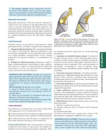
                                                               Figure 17.13  Common locations of lung abscess. A, Primary lung
           Lipid Pneumonia                                     abscess—mostly single, large, commonly due to aspiration, located most
                                                               frequently in the lower part of right upper lobe or apex of right lower lobe.
           Another variety of non-infective pneumonia is lipid  B, Secondary lung abscesses—mostly multiple, small, most commonly
           pneumonia which is of 2 types: exogenous and endogenous.  post-pneumonic or following septic embolism.
           1. Exogenous lipid pneumonia. This is caused by aspiration
           of a variety of oily materials. These are: inhalation of oily  are introduced into the lungs from one of the following
           nasal drops, regurgitation of oily medicines from stomach  mechanisms:
           (e.g. liquid paraffin), administration of oily vitamin  1. Aspiration of infected foreign material. A number of
           preparation to reluctant children or to debilitated old  foreign materials such as food, decaying teeth, gastric
           patients.                                           contents, severely infected gingivae and teeth, and necrotic  CHAPTER 17
           2. Endogenous lipid pneumonia. Endogenous origin of  tissue from lesions in the mouth, upper respiratory tract or
           lipids causing pneumonic consolidation is more common.  nasopharynx may be aspirated. This occurs particularly in
           The sources of origin are tissue breakdown following  favourable circumstances such as during sleep,
           obstruction to airways e.g. obstruction by bronchogenic  unconsciousness, anaesthesia, general debility and acute
           cancer, tuberculosis and bronchiectasis.            alcoholism.
                                                               2. Preceding bacterial infection. Preceding broncho-
            MORPHOLOGIC FEATURES. Grossly, the exogenous       pneumonia in a debilitated patient may develop into lung
            lipid pneumonia affects the right lung more frequently  abscess. Other infective conditions like tuberculosis,
            due to direct path from the main bronchus. Quite often,  bronchiectasis and mycotic infections may occasionally result
            the lesions are bilateral. The affected part of the lungs is  in formation of lung abscess.
            consolidated. Cut surface is characteristically ‘golden  3. Bronchial obstruction. An abscess may form distal to an  The Respiratory System
            yellow’.                                           obstructed bronchus such as from bronchial tumour or from
            Microscopically, the features are as under:        impacted foreign body.
            i) Lipid is finely dispersed in the cytoplasm of
            macrophages forming foamy macrophages within the   4. Septic embolism. Infected emboli originating from
            alveolar spaces.                                   pyaemia, thrombophlebitis or from vegetative bacterial
            ii) There may be formation of cholesterol clefts due to  endocarditis may be disseminated in the venous circulation
            liberation of cholesterol and other lipids.        and reach the right side of the heart from where they are
            iii) Formation of granulomas with foreign body giant cells  lodged in the lung and result in multiple abscesses.
            may be seen around the large lipid droplets.       5. Miscellaneous. Rarely lung abscesses may occur from
                                                               following causes:
           LUNG ABSCESS                                        i) Infection in pulmonary infarcts.
                                                               ii) Amoebic abscesses due to infection with  Entamoeba
           Lung abscess is a localised area of necrosis of lung tissue  histolytica.
           with suppuration. It is of 2 types (Fig. 17.13):    iii) Trauma to the lungs.
              Primary lung abscess that develops in an otherwise normal  iv) Direct extension from a suppurative focus in the
           lung. The commonest cause is aspiration of infected material.  mediastinum, oesophagus, subphrenic area or spine.
              Secondary lung abscess that develops as a complication of  MORPHOLOGIC FEATURES. Abscesses due to aspira-
           some other disease of the lung or from another site.  tion are more likely to be in right lung due to more vertical
                                                                 main bronchus and are frequently single. They are
           ETIOPATHOGENESIS. The microorganisms commonly         commonly located in the lower part of the right upper
           isolated from the lungs in lung abscess are streptococci,  lobe or apex of right lower lobe. Abscesses developing
           staphylococci and various gram-negative organisms. These

