


Page 515 - Textbook of Pathology, 6th Edition
P. 515
or amplification of the EGFR gene. EGFR belonging to ERBB 499
(HER) family of protooncogenes through mutation in its
tyrosine kinase (TK) domain plays a role in both extracelluar
and intracellular signaling resulting in tumour cell
proliferation, metastasis and antiapoptotic action. Targeted
molecular therapy against these mutations in EGFR include
EGFR-TK inhibitor oral therapy.
ii) VEGF and monoclonal therapy: Although not mutated, VEGF
is excessively produced in lung cancer and contributes to
tumour angiogenesis. Monoclonal antibody therapy against
EGFR in conjunction with chemotherapy has been used for
curtailing tumour angiogenesis in lung caner.
iii) Molecular signature gene for prediction: Recent proteomic
studies at research level have shown that each patient has
unique protein pattern in the serum (i.e. molecular
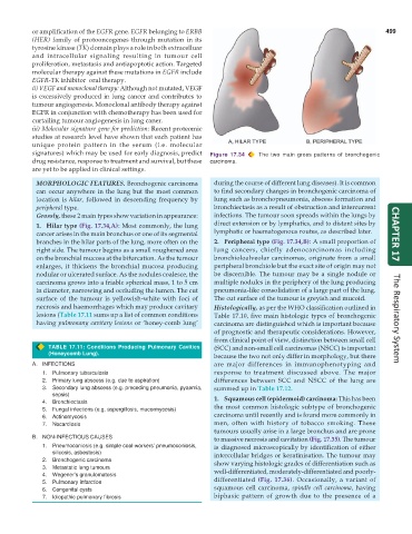
signatures) which may be used for early diagnosis, predict Figure 17.34 The two main gross patterns of bronchogenic
drug resistance, response to treatment and survival, but these carcinoma.
are yet to be applied in clinical settings.
MORPHOLOGIC FEATURES. Bronchogenic carcinoma during the course of different lung diseases). It is common
can occur anywhere in the lung but the most common to find secondary changes in bronchogenic carcinoma of
location is hilar, followed in descending frequency by lung such as bronchopneumonia, abscess formation and
peripheral type. bronchiectasis as a result of obstruction and intercurrent
Grossly, these 2 main types show variation in appearance: infections. The tumour soon spreads within the lungs by
1. Hilar type (Fig. 17.34,A): Most commonly, the lung direct extension or by lymphatics, and to distant sites by
cancer arises in the main bronchus or one of its segmental lymphatic or haematogenous routes, as described later. CHAPTER 17
branches in the hilar parts of the lung, more often on the 2. Peripheral type (Fig. 17.34,B): A small proportion of
right side. The tumour begins as a small roughened area lung cancers, chiefly adenocarcinomas including
on the bronchial mucosa at the bifurcation. As the tumour bronchioloalveolar carcinomas, originate from a small
enlarges, it thickens the bronchial mucosa producing peripheral bronchiole but the exact site of origin may not
nodular or ulcerated surface. As the nodules coalesce, the be discernible. The tumour may be a single nodule or
carcinoma grows into a friable spherical mass, 1 to 5 cm multiple nodules in the periphery of the lung producing
in diameter, narrowing and occluding the lumen. The cut pneumonia-like consolidation of a large part of the lung.
surface of the tumour is yellowish-white with foci of The cut surface of the tumour is greyish and mucoid.
necrosis and haemorrhages which may produce cavitary Histologically, as per the WHO classification outlined in
lesions (Table 17.11 sums up a list of common conditions Table 17.10, five main histologic types of bronchogenic
having pulmonany cavitary lesions or ‘honey-comb lung’ carcinoma are distinguished which is important because The Respiratory System
of prognostic and therapeutic considerations. However,
from clinical point of view, distinction between small cell
TABLE 17.11: Conditions Producing Pulmonary Cavities (SCC) and non-small cell carcinomas (NSCC) is important
(Honeycomb Lung). because the two not only differ in morphology, but there
A. INFECTIONS are major differences in immunophenotyping and
1. Pulmonary tuberculosis response to treatment discussed above. The major
2. Primary lung abscess (e.g. due to aspiration) differences between SCC and NSCC of the lung are
3. Secondary lung abscess (e.g. preceding pneumonia, pyaemia, summed up in Table 17.12.
sepsis) 1. Squamous cell (epidermoid) carcinoma: This has been
4. Bronchiectasis
5. Fungal infections (e.g. aspergillosis, mucormycosis) the most common histologic subtype of bronchogenic
6. Actinomycosis carcinoma until recently and is found more commonly in
7. Nocardiosis men, often with history of tobacco smoking. These
tumours usually arise in a large bronchus and are prone
B. NON-INFECTIOUS CAUSES to massive necrosis and cavitation (Fig. 17.35). The tumour
1. Pneumoconiosis (e.g. simple coal-workers’ pneumoconiosis, is diagnosed microscopically by identification of either
silicosis, asbestosis) intercellular bridges or keratinisation. The tumour may
2. Bronchogenic carcinoma show varying histologic grades of differentiation such as
3. Metastatic lung tumours well-differentiated, moderately-differentiated and poorly-
4. Wegener’s granulomatosis
5. Pulmonary infarction differentiated (Fig. 17.36). Occasionally, a variant of
6. Congenital cysts squamous cell carcinoma, spindle cell carcinoma, having
7. Idiopathic pulmonary fibrosis biphasic pattern of growth due to the presence of a

