


Page 519 - Textbook of Pathology, 6th Edition
P. 519
ii) Other systemic manifestations:  These include the                                                    503
           following:
           a) Neuromuscular e.g. polymyositis, myopathy, peripheral
           neuropathy and subacute cerebellar degeneration.
           b) Skeletal e.g. clubbing and hypertrophic osteoarthro-
           pathy.
           c) Cutaneous e.g. acanthosis nigricans and dermato-
           myopathy.
           d) Cardiovascular e.g. migratory thrombophlebitis
           (Trousseau’s syndrome), nonbacterial thrombotic
           endocarditis.
           e) Haematologic e.g. abnormalities in coagulation and
           leukaemoid reaction.

           STAGING AND PROGNOSIS. The widely accepted clinical
           staging of lung cancer is according to the TNM classification,
           combining features of primary Tumours, Nodal involvement
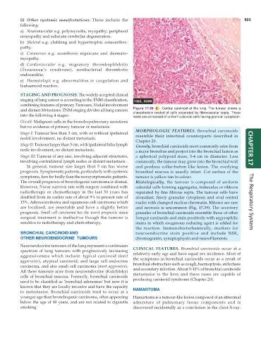
           and distant Metastases. TNM staging divides all lung cancers  Figure 17.39  Central carcinoid of the lung. The tumour shows a
           into the following 4 stages:                        characteristic nested of cells separated by fibrovascular septa. These
                                                               nests are composed of uniform cuboidal cells having granular cytoplasm.
           Occult: Malignant cells in the bronchopulmonary secretions
           but no evidence of primary tumour or metastasis.
                                                                 MORPHOLOGIC FEATURES. Bronchial carcinoids
           Stage I: Tumour less than 3 cm, with or without ipsilateral
           nodal involvement, no distant metastasis.             resemble their intestinal counterparts described in
                                                                 Chapter 20.
           Stage II: Tumour larger than 3 cm, with ipsilateral hilar lymph  Grossly, bronchial carcinoids most commonly arise from
           node involvement, no distant metastasis.              a major bronchus and project into the bronchial lumen as  CHAPTER 17
           Stage III: Tumour of any size, involving adjacent structures,  a spherical polypoid mass, 3-4 cm in diameter. Less
           involving contralateral lymph nodes or distant metastasis.  commonly, the tumour may grow into the bronchial wall
              In general, tumour size larger than 5 cm has worse  and produce collar-button like lesion. The overlying
           prognosis. Symptomatic patients, particularly with systemic  bronchial mucosa is usually intact. Cut surface of the
           symptoms, fare far badly than the nonsymptomatic patients.  tumour is yellow-tan in colour.
           The overall prognosis of bronchogenic carcinoma is dismal.  Histologically, the tumour is composed of uniform
           However, 5-year survival rate with surgery combined with  cuboidal cells forming aggregates, trabeculae or ribbons
           radiotherapy or chemotherapy in the last 30 years has  separated by fine fibrous septa. The tumour cells have
           doubled from its earlier rate of about 9% to present rate of  abundant, finely granular cytoplasm and oval central
           15%. Adenocarcinoma and squamous cell carcinoma which  nuclei with clumped nuclear chromatin. Mitoses are rare  The Respiratory System
           are localised, are resectable and have a slightly better  and necrosis is uncommon (Fig. 17.39). The secretory
           prognosis. Small cell carcinoma has the worst prognosis since  granules of bronchial carcinoids resemble those of other
           surgical treatment is ineffective though the tumour is  foregut carcinoids and stain positively with argyrophilic
           sensitive to radiotherapy and chemotherapy.           stains in which exogenous reducing agent is added for
                                                                 the reaction. Immunohistochemically, markers for
           BRONCHIAL CARCINOID AND                               neuroendocrine stain positive and include NSE,
           OTHER NEUROENDOCRINE  TUMOURS                         chromogranin, synaptophysin and neurofilaments.
           Neuroendocrine tumours of the lung represent a continuum  CLINICAL FEATURES.  Bronchial carcinoids occur at a
           spectrum of lung tumours with progressively increasing  relatively early age and have equal sex incidence. Most of
           aggressiveness which include: typical carcinoid (least  the symptoms in bronchial carcinoids occur as a result of
           aggressive), atypical carcinoid, and large cell endocrine  bronchial obstruction such as cough, haemoptysis, atelectasis
           carcinoma, and also small cell carcinoma (most aggressive).  and secondary infection. About 5-10% of bronchial carcinoids
           All these tumours arise from neuroendocrine (Kulchitsky)  metastasise to the liver and these cases are capable of
           cells of bronchial mucosa. Formerly, bronchial carcinoids  producing carcinoid syndrome (Chapter 20).
           used to be classified as ‘bronchial adenomas’ but now it is
           known that they are locally invasive and have the capacity
           to metastasise. Bronchial carcinoids tend to occur at a  HAMARTOMA
           younger age than bronchogenic carcinoma, often appearing  Hamartoma is a tumour-like lesion composed of an abnormal
           below the age of 40 years, and are not related to cigarette  admixture of pulmonary tissue components and is
           smoking.                                            discovered incidentally as a coin-lesion in the chest-X-ray.

