


Page 520 - Textbook of Pathology, 6th Edition
P. 520
504 Pulmonary hamartomas are of 2 types: chondromatous and                    PLEURA
           leiomyomatous.
              Chondromatous hamartoma is more common and       NORMAL STRUCTURE
           usually asymptomatic. It forms a solitary, spherical mass,  Visceral pleura covers the lungs and extends into the fissures
           2-5 cm in diameter, usually at the periphery of the lung.  while the parietal pleura limits the mediastinum and covers
           Typically, it shows nodules of cartilage associated with  the dome of the diaphragm and inner aspect of the chest wall.
           fibrous and adipose tissue admixed with bronchial   The two layers between them enclose pleural cavity which
           epithelium.
                                                               contains less than 15 ml of clear serous fluid.
              Leiomyomatous hamartoma has a prominent smooth
           muscle component and bronchiolar structures. They are  Microscopically, both the pleural layers are lined by a single
           frequently multiple, 1-2 mm in diameter and are more  layer of flattened mesothelial cells facing each other.
           commonly located near the pleura.                   Underneath the lining cells is a thin layer of connective tissue.
                                                                  Diseases affecting the pleura are nearly always secondary
                                                               to some other underlying disease. Broadly, they fall into
           METASTATIC LUNG TUMOURS
                                                               inflammations, non-inflammatory pleural effusions,
           Secondary tumours of the lungs are more common than the  pneumothorax, and tumours.
           primary pulmonary tumours. Metastases from carcinomas
           as well as sarcomas arising from anywhere in the body may  INFLAMMATIONS
           spread to the lung by haematogenous or lymphatic routes,  Inflammatory involvement of the pleura is commonly termed
           or by direct extension. Blood-borne metastases are the most  pleuritis or pleurisy. Depending upon the character of resultant
           common since emboli of tumour cells from any malignant  exudate, it can be divided into serous, fibrinous and
           tumour entering the systemic venous circulation are likely  serofibrinous, suppurative or empyema, and haemorrhagic
           to be lodged in the lungs. Metastases are most common in  pleuritis.
           the peripheral part of the lung forming single or multiple,
           discrete nodular lesions which appear radiologically as  1. SEROUS, FIBRINOUS AND SEROFIBRINOUS
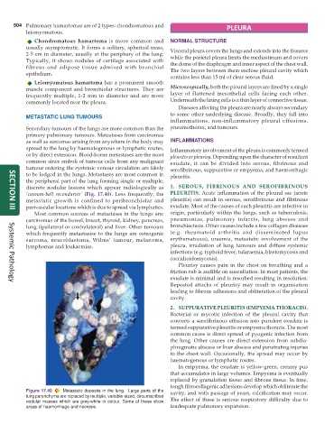
           ‘cannon-ball secondaries’  (Fig. 17.40). Less frequently, the  PLEURITIS. Acute inflammation of the pleural sac (acute
           metastatic growth is confined to peribronchiolar and  pleuritis) can result in serous, serofibrinous and fibrinous
           perivascular locations which is due to spread via lymphatics.  exudate. Most of the causes of such pleuritis are infective in
     SECTION III
              Most common sources of metastases in the lungs are:  origin, particularly within the lungs, such as tuberculosis,
           carcinomas of the bowel, breast, thyroid, kidney, pancreas,  pneumonias, pulmonary infarcts, lung abscess and
           lung (ipsilateral or contralateral) and liver. Other tumours  bronchiectasis. Other causes include a few collagen diseases
           which frequently metastasise to the lungs are osteogenic  (e.g. rheumatoid arthritis and disseminated lupus
           sarcoma, neuroblastoma, Wilms’ tumour, melanoma,    erythematosus), uraemia, metastatic involvement of the
           lymphomas and leukaemias.                           pleura, irradiation of lung tumours and diffuse systemic
                                                               infections (e.g. typhoid fever, tularaemia, blastomycosis and
                                                               coccidioidomycosis).
                                                                  Pleurisy causes pain in the chest on breathing and a
                                                               friction rub is audible on auscultation. In most patients, the
                                                               exudate is minimal and is resorbed resulting in resolution.
                                                               Repeated attacks of pleurisy may result in organisation
     Systemic Pathology
                                                               leading to fibrous adhesions and obliteration of the pleural
                                                               cavity.

                                                               2. SUPPURATIVE PLEURITIS (EMPYEMA THORACIS).
                                                               Bacterial or mycotic infection of the pleural cavity that
                                                               converts a serofibrinous effusion into purulent exudate is
                                                               termed suppurative pleuritis or empyema thoracis. The most
                                                               common cause is direct spread of pyogenic infection from
                                                               the lung. Other causes are direct extension from subdia-
                                                               phragmatic abscess or liver abscess and penetrating injuries
                                                               to the chest wall. Occasionally, the spread may occur by
                                                               haematogenous or lymphatic routes.
                                                                  In empyema, the exudate is yellow-green, creamy pus
                                                               that accumulates in large volumes. Empyema is eventually
                                                               replaced by granulation tissue and fibrous tissue. In time,
                                                               tough fibrocollagenic adhesions develop which obliterate the
           Figure 17.40  Metastatic deposits in the lung.  Large parts of the  cavity, and with passage of years, calcification may occur.
           lung parenchyma are replaced by multiple, variable-sized, circumscribed  The effect of these is serious respiratory difficulty due to
           nodular masses which are grey-white in colour. Some of these show
           areas of haemorrhage and necrosis.                  inadequate pulmonary expansion.

