


Page 516 - Textbook of Pathology, 6th Edition
P. 516
500
TABLE 17.12: Comparison of Features of Small Cell and Non-small Cell Carcinoma of the Lung.
Feature Small Cell Carcinoma Non-small Cell Carcinoma
1. Etiologic relationship Strongly related to tobacco smoking Smoking implicated, other factors:
pollution, chronic scars, asbestos exposure
2. Morphology
i) Pattern Diffuse sheets Squamous or glandular pattern
ii) Nuclei Hyperchromatic, fine chromatin Pleomorphic, coarse chromatin
iii) Nucleoli Indistinct Prominent
iv) Cytoplasm Scanty Abundant
3. Neuroendocrine markers Present Absent
(e.g. dense-core granules on EM,
chromogranin, synaptophysin,
neuron-specific enolase, CD56, CD57)
4. Epithelial markers Present Present
(e.g. epithelial membrane antigen,
carcinoembryonic antigen, cytokeratin)
5. Mucin Absent Present in adenocarcinoma
6. HLA, β2 microglobulin Absent to low Present
7. Peptide hormone production Gastrin, ACTH, ADH, calcitonin Parathormone
8. Genetic abnormalities 3p allele loss, RB and 3p allele loss, EGFR and K-RAS
p53 mutations mutations
9. Treatment type Radiotherapy and/or Surgical resection possible, limited
chemotherapy response to radiotherapy and/or
chemotherapy
10. Prognosis Poor Better
component of squamous cell carcinoma and the other bronchi show squamous metaplasia, epithelial dysplasia
SECTION III
sarcoma-like spindle cell component, is found. and carcinoma in situ.
Usually the spread of squamous cell carcinoma is more 2. Small cell carcinoma: Small cell carcinomas are
rapid than the other histologic types of NSCC. Frequently, frequently hilar or central in location, have strong
the edge of the growth and the adjoining uninvolved relationship to cigarette smoking and are highly malignant
tumours. They are most often associated with ectopic
hormone production because of the presence of
neurosecretory granules in majority of tumour cells which
are similar to those found in argentaffin or Kulchitsky cells
normally found in bronchial epithelium. By immuno-
histochemistry, these tumour cells are positive for
neuroendocrine markers: chromogranin, neuron-specific
Systemic Pathology
enolase (NSE) and synaptophysin. Small cell carcinomas
have 3 subtypes:
i) Oat cell carcinoma is composed of uniform, small cells,
larger than lymphocytes with dense, round or oval nuclei
having diffuse chromatin, inconspicuous nucleoli and very
sparse cytoplasm (oat = a form of grain). These cells are
organised into cords, aggregates and ribbons or around
small blood vessels forming pseudorosettes (Fig. 17.37).
ii) Small cell carcinoma, intermediate cell type is composed of
cells slightly larger than those of oat cell carcinoma and
have similar nuclear characteristics but have more
abundant cytoplasm. These cells are organised into lobules.
iii) Combined oat cell carcinoma is a tumour in which there
is a definite component of oat cell carcinoma with
squamous cell and/or adenocarcinoma.
3. Adenocarcinoma: Adenocarcinoma, also called
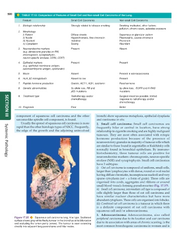
Figure 17.35 Squamous cell carcinoma lung, hilar type. Sectioned peripheral carcinoma due to its location and scar carcinoma
surface shows grey-white fleshy tumour in the bronchus at its bifurcation due to its association with areas of chronic scarring, is the
and occluding the lumen partly (arrow). The tumour is seen extending
directly into adjacent lung parenchyma and hilar nodes. most common bronchogenic carcinoma in women and is

