


Page 517 - Textbook of Pathology, 6th Edition
P. 517
501


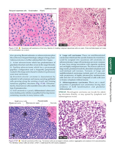
           Figure 17.36  Squamous cell carcinoma of the lung. Islands of invading malignant squamous cells are seen. A few well-developed cell nests
           with keratinisation are evident.


            slow-growing. Recent estimates on adenocarcinoma place  4. Large cell carcinoma: These are undifferentiated
            this as the most frequent histologic subtype of lung cancer.  carcinomas which lack the specific features by which they
            Adenocarcinoma is further subclassified into 4 types:  could be assigned into squamous cell carcinoma or
            i) Acinar adenocarcinoma which has predominance of   adenocarcinoma. Large cell carcinomas are more common
            glandular structure and often occurs in the larger bronchi.  in men, have strong association with cigarette smoking
            ii) Papillary adenocarcinoma which has a pronounced  and are highly malignant tumours. The tumour cells have  CHAPTER 17
            papillary configuration and is frequently peripherally  large nuclei, prominent nucleoli, abundant cytoplasm and
            located in the lungs and is found in relation to pulmonary  well-defined cell borders. Variants of large cell
            scars (scar carcinoma).                              undifferentiated carcinomas include giant cell carcinoma
                                                                 with prominence of highly pleomorphic multinucleate
            iii) Bronchiolo-alveolar carcinoma is characterised by  cells and clear cell carcinoma composed of cells with clear
            cuboidal to tall columnar and mucus-secreting epithelial  or foamy cytoplasm without mucin.
            cells growing along the existing alveoli and forming  5. Adenosquamous carcinoma: These are a small
            numerous papillary structures  (Fig. 17.38). Ultrastruc-  proportion of peripheral scar carcinomas having clear
            turally, these tumour cells resemble Clara cells or less often  evidence of both keratinisation and glandular
            type II pneumocytes.                                 differentiation.
            iv) Solid carcinoma is a poorly-differentiated adenocarci-                                                The Respiratory System
            noma lacking acini, tubules or papillae but having mucus-  SPREAD. Bronchogenic carcinoma can invade the adjoin-
            containing vacuoles in many tumour cells.          ing structures directly, or may spread by lymphatic and
                                                               haematogenous routes.


           Figure 17.37  Oat cell carcinoma of the lung. The tumour cells are arranged in sheets, cords or aggregates and at places form pseudorosettes.
           The individual tumour cells are small, uniform, lymphocyte-like with scanty cytoplasm.

