
Page 640 - Textbook of Pathology, 6th Edition
P. 640
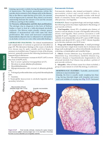
624                                                      Post-necrotic Cirrhosis
            forming regenerative nodules having disorganised masses
            of hepatocytes. The hepatic parenchyma within the  Post-necrotic cirrhosis, also termed  post-hepatitic cirrhosis,
            nodules shows extensive fatty change early in the disease.  macronodular cirrhosis and  coarsely nodular cirrhosis, is
            But as the fibrous septa become more thick, the amount  characterised by large and irregular nodules with broad
            of fat in hepatocytes is reduced. Thus, there is an inverse  bands of connective tissue and occurring most commonly
            relationship between the amount of fat and the amount  after previous viral hepatitis.
            of fibrous scarring in the nodules.                ETIOLOGY. Based on epidemiologic and serologic studies,
            iv) Necrosis, inflammation and bile duct proliferation:  the following factors have been implicated in the etiology of
            The etiologic clue to diagnosis in the form of Mallory  post-necrotic cirrhosis.
            bodies is hard to find in a fully-developed alcoholic  1. Viral hepatitis. About 25% of patients give history of
            cirrhosis. The fibrous septa usually contain sparse  recent or remote attacks of acute viral hepatitis followed by
            infiltrate of mononuclear cells with some bile duct  chronic viral hepatitis. Most common association is with
            proliferation. Bile stasis and increased cytoplasmic  hepatitis B and C; hepatitis A is not known to evolve into
            haemosiderin deposits due to enhanced iron absorption
            in alcoholic cirrhosis are some other noticeable findings.  cirrhosis. It is estimated that about 20% cases of HBV chronic
                                                               hepatitis and about 20-30% cases of HCV chronic hepatitis
           LABORATORY DIAGNOSIS. The clinical manifestations   go to develop cirrhosis over 20-30 years.
           and complications of cirrhosis in general are described on  2. Drugs and chemical hepatotoxins. A small percentage
           page 630. The laboratory findings in the course of alcoholic  of cases may have origin from toxicity due to chemicals and
           liver disease may be quite variable and liver biopsy is  drugs such as phosphorus, carbon tetrachloride, mushroom
           necessary in doubtful cases. Progressive form of the disease,  poisoning, acetaminophen and α-methyl dopa.
           however, generally presents the following biochemical and  3. Others. Certain infections (e.g. brucellosis), parasitic
           haematological alterations:                         infestations (e.g. clonorchiasis), metabolic diseases (e.g.
           1. Elevated transaminases: increase in SGOT (AST) is more  Wilson’s disease or hepatolenticular degeneration) and
           than that of SGPT (ALT).                            advanced alcoholic liver disease may produce a picture of
           2. Rise in serum γ-glutamyl transpeptidase (γ-GT).  post-necrotic cirrhosis.
           3. Elevation in serum alkaline phosphatase.         4. Idiopathic. After all these causes have been excluded, a
           4. Hyperbilirubinaemia.                             group of cases remain in which the etiology is unknown.
           5. Hypoproteinaemia with reversal of albumin-globulin
     SECTION III
           ratio.                                                MORPHOLOGIC FEATURES. Typically, post-necrotic
           6. Prolonged prothrombin time and partial thromboplastin  cirrhosis is macronodular type.
           time.                                                 Grossly, the liver is usually small, weighing less than
           7. Anaemia.                                           1 kg, having distorted shape with irregular and coarse
           8. Neutrophilic leucocytosis in alcoholic hepatitis and in  scars and nodules of varying size (Fig. 21.27). Sectioned
           secondary infections.


     Systemic Pathology


           Figure 21.27  Post-necrotic cirrhosis, showing the typical irregular
           macronodular pattern (nodules larger than 3 mm diameter). Externally
           the liver is small, distorted and irregularly scarred.